7 – 9 April 2025
Imaging Methods Core Facility, BIOCEV, Průmyslová 595, 252 50, Vestec
The course will focus on basic operations in cryogenic electron microscopy (cryoEM), cryoimaging of vitrified particulate specimens (e.g. microtubules, vesicles, viruses, etc.) and the possibility of correlation with cryogenic fluorescence microscopy (cryoFM). The lectures will cover the theoretical principles of EM/cryoEM and FM/cryoFM. During hands-on sessions, participants will gain practical experience with cryo-sample preparation techniques (Plunge Freezer, High Pressure Freezing) and cryo-imaging using confocal fluorescence microscope and transmission electron microscope. The course is particularly suitable for beginners in cryoEM with little or no experience in the field.

31 March – 4 April 2025
Light Microscopy Core Facility, Institute of Molecular Genetics, Vídeňská 1083, 142 20, Prague
The course will address fundamental aspects of image data acquisition, processing, and analysis, encompassing techniques in stereology. Alongside theoretical principles, the course will prioritize hands-on practical learning. Participants will gain proficiency in utilizing the freely available software package Fiji for both basic and advanced analyses. They will learn to assess co-localizations, analyze data from FRAP and electron microscopy, track particles, segment objects in images, and explore methods of employing artificial intelligence for image segmentation.
Additionally, participants will master techniques to enhance data quality through deconvolution using Huygens software. An interactive session featuring Imaris software will be complemented by practical exercises. Furthermore, this year introduces new demos for analyses using the NIS Elements package and a lecture about the proper way of publishing images in scientific journals. Independent practical tasks involving Fiji and Huygens will also constitute a significant component of the course.
While the course aligns with the Microscopy Methods in Biomedicine, prior attendance is not a prerequisite for participation.

3-7 March 2025
Electron Microscopy Core Facility, Institute of Molecular Genetics, Vídeňská 1083, 142 20, Prague
The course is designed for users of electron microscopes in biomedical field, both from scientific laboratories and from core facilities. The aim of the course is to provide an orientation to basic and special techniques in biomedical electron microscopy in an application-oriented manner. In the theroretical and practical sessions, sample preparation under room temperature and cryo conditions will be covered, including special workflows such as cryo-sectioning, freeze-fracture replica, or cryo-CLEM workflow on FIB-milled lamella. Besides basic imaging, special techniques such as (cryo)FIB-SEM, STEM tomography and EDS analysis will be demonstrated, with introduction to data processing and interpretation. Special emphasis will be placed on discussing how to choose the appropriate methodology for a specific scientific project.
28-30 May 2025
Imaging Methods Core Facility, BIOCEV, Průmyslová 595, 252 50, Vestec
The fastest and most gentle 3D live-cell imaging on the Carl Zeiss Lattice Lightsheet 7, used for tracking the trafficking of fluorescently labeled proteins in living cells, will be complemented by FRAP on the Nikon iLas 2 ring-TIRF/FRAP and Nikon CSU-W1, as well as by FCS on the Leica TCS SP8 WLL SMD-FLIM for measuring protein diffusion in the plasma membrane of living cells. Finally, model systems of molecular motors walking along microtubules, visualized using the Nikon Ti-E H-TIRF, will demonstrate the benefits of single-molecule approaches for studying molecular motion.
This course includes theoretical and hands-on training in four complementary microscopy approaches for visualizing and measuring the lateral movement of molecules in living cells or model systems.
The microscopy techniques covered in the course:
11-12 February 2025
Building Da I. IPHYS BIF, Krc Campus, Videnska 1083, Prague
The two-day course consists of lectures and hands-on sessions which will demonstrate basics in design of experiments, for instance hypothesizing, sampling, data dependency, statistical power or hypothesis testing in biology. The examples will be provided in the field of light microscopy and proteomics. The participants will learn the ways to correctly acquire data using high-end microscopes using a wide pallet of methods such as FLIM, SHG or CARS or proteomic approaches. In addition to that the participants will process and analyse the data using traditional approaches, stereology or AI tools or VR in Fiji or Python. Finally, the participants will be taught the data interpretation and presentation.
Although the course is biology science oriented, the skills you will acquire are valid and necessary in any area of research, development or industry.

3-7 February 2025
Room B311, Viničná Microscopy Core Facility, Viničná 1594/7, 128 00 Prague
This 5-day workshop is intended to give a thorough introduction to Bioimage analysis for individuals with little to no prior experience but who want to develop some basic skills in image processing and analysis workflows using both open-source and proprietary software. Participants will get hands-on experience with image restoration, segmentation techniques, feature extraction and tracking using classical and advanced machine and deep learning tools. Most importantly, students will gain insight in data visualization and analysis for reproducible research. By the end of the workshop, attendees will have the opportunity to work on a final project, in which they will be able to apply the newly learned skills on a real-world bioimage analysis challenge.
By the end of this workshop, participants will be able to:
27-31 January 2025
Viničná Microscopy Core Facility, Viničná 1594/7, 128 00 Prague
Join the comprehensive course organized by the Viničná Microscopy Core Facility at the Charles University, Prague to master image analysis using cutting-edge tools like ImageJ, Huygens, Arivis Vision4D, and Python. You’ll learn key techniques including deconvolution, segmentation, colocalization, and deep learning models, alongside practical applications in biological imaging.
Course Highlights:
Perfect for researchers and scientists looking to elevate their image analysis skills!
Course website:
To sign up contact the course organizer Mgr. Zuzana Burdíková, Ph.D. at burdika@natur.cuni.cz
Dear Czech-BioImaging users,
we would like to share with you the announcement of the Wellcome Trust.
The Wellcome Photography Prize is back and accepting entries once again, after being on pause since the pandemic. Excitingly, scientific and medical imaging images are accepted once again. The unique view that scientific and medical imaging can reveal will add a really interesting perspective to the photography alongside which it will sit.
The Wellcome Photography Prize returns to celebrate captivating stories of health, science and human experience, bringing together different perspectives from around the world. Free to enter and open to all, entrants could win one of 25 prizes up to £10,000, have their work shown in a major public exhibition and receive international media coverage. Don’t miss the chance to share a story you’re passionate about. Entries close 14 January 2025.
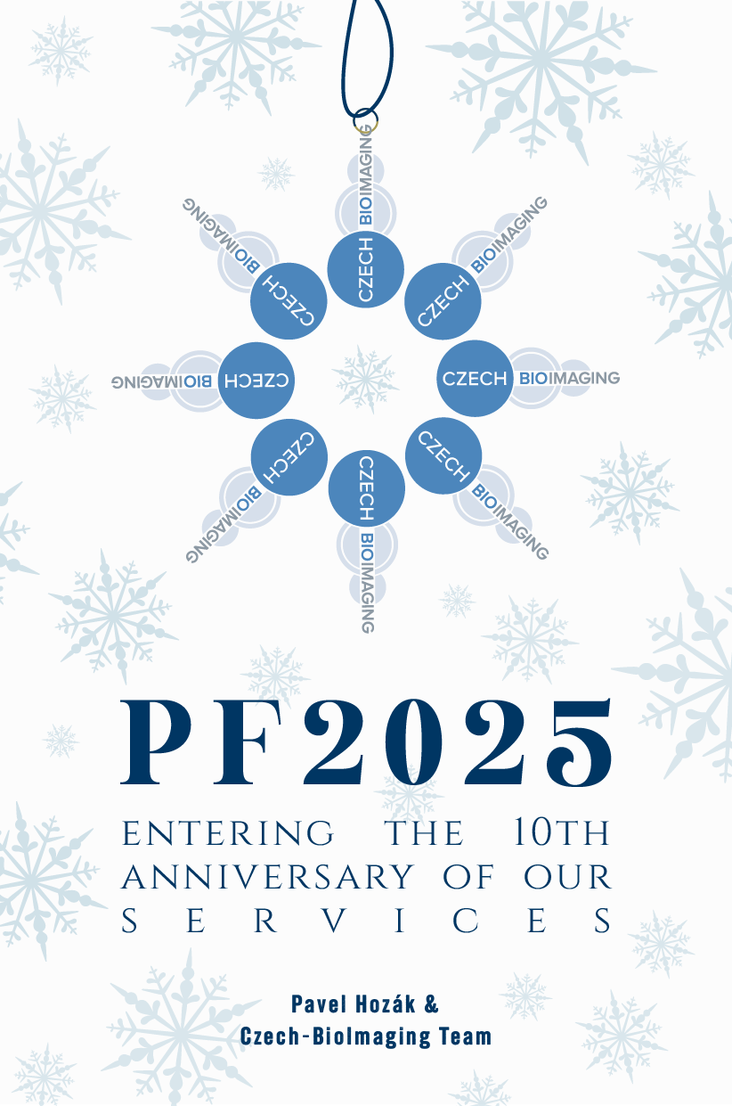
Dear Colleagues and Friends,
as 2024 draws to a close, we would like to take a moment to wish you happy holidays.
We deeply appreciate your trust, enthusiasm, and the excellent research projects and results made possible through your use of Czech-BioImaging. Your inspiring work and collaborations motivate us to continuously improve and grow as a research infrastructure.
As we enter 2025, a year in which Czech-BioImaging celebrates 10 years of advancing biological and medical imaging, we remain committed to providing the highest quality imaging services to support your research endeavors.
Wishing you much success, both professional and personal, in the year ahead!
Dear Czech-BioImaging users,
we would like to share with you the announcement for the Ernst-Ruska-Prize 2025 of the German Society for Electron Microscopy.
The submission deadline is November 30th, 2024.
PDF: http://www.dge-homepage.de/erp2025_announcement.pdf
More information is available at: http://www.dge-homepage.de/
Please also note the announcement of the Harald Rose Distinguished Lecture Award 2025 for which the deadline is February 28th, 2025.
See also at http://www.dge-homepage.de/hrl2025_announcement.pdf


Dear Czech-BioImaging users,
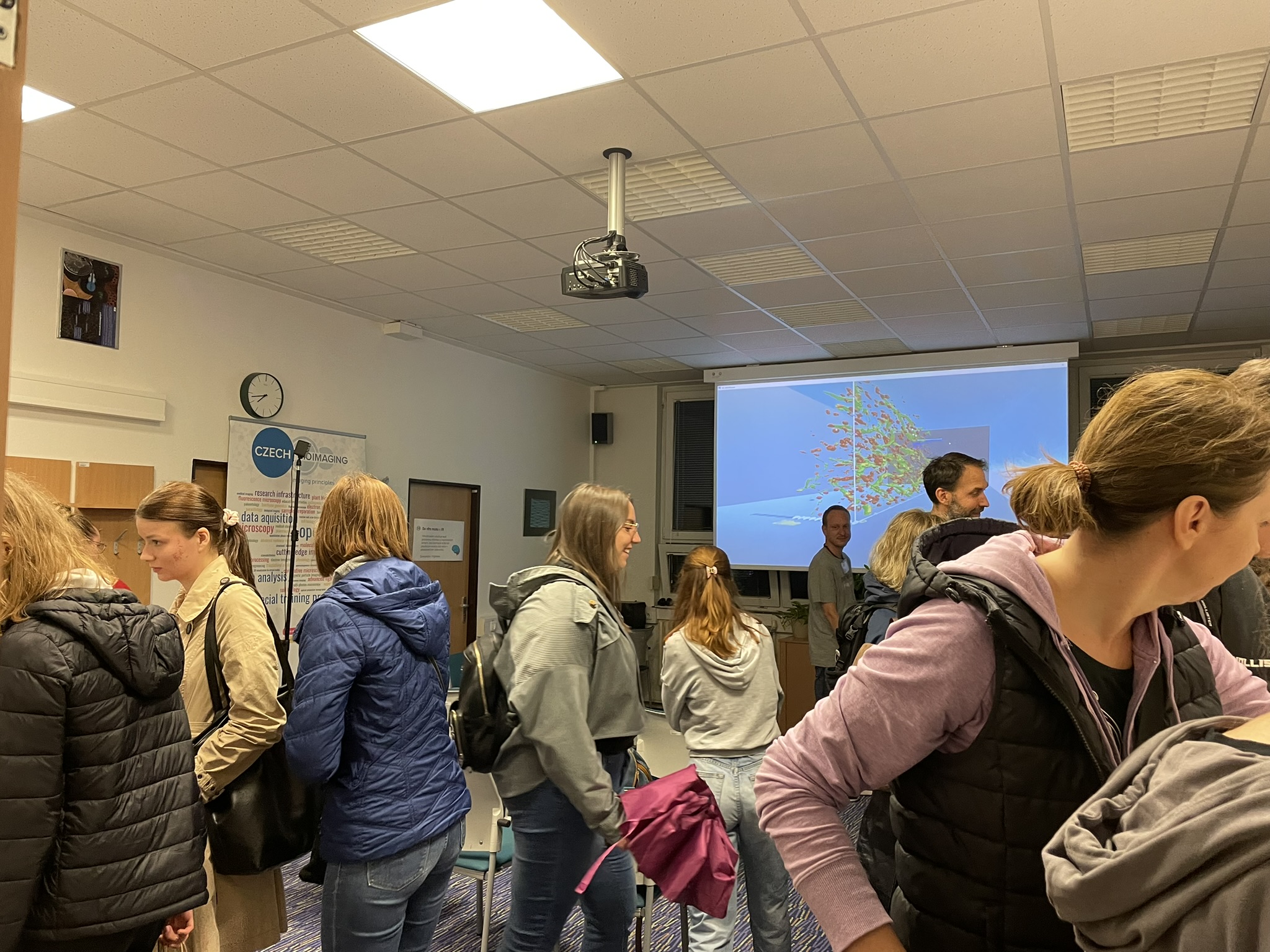
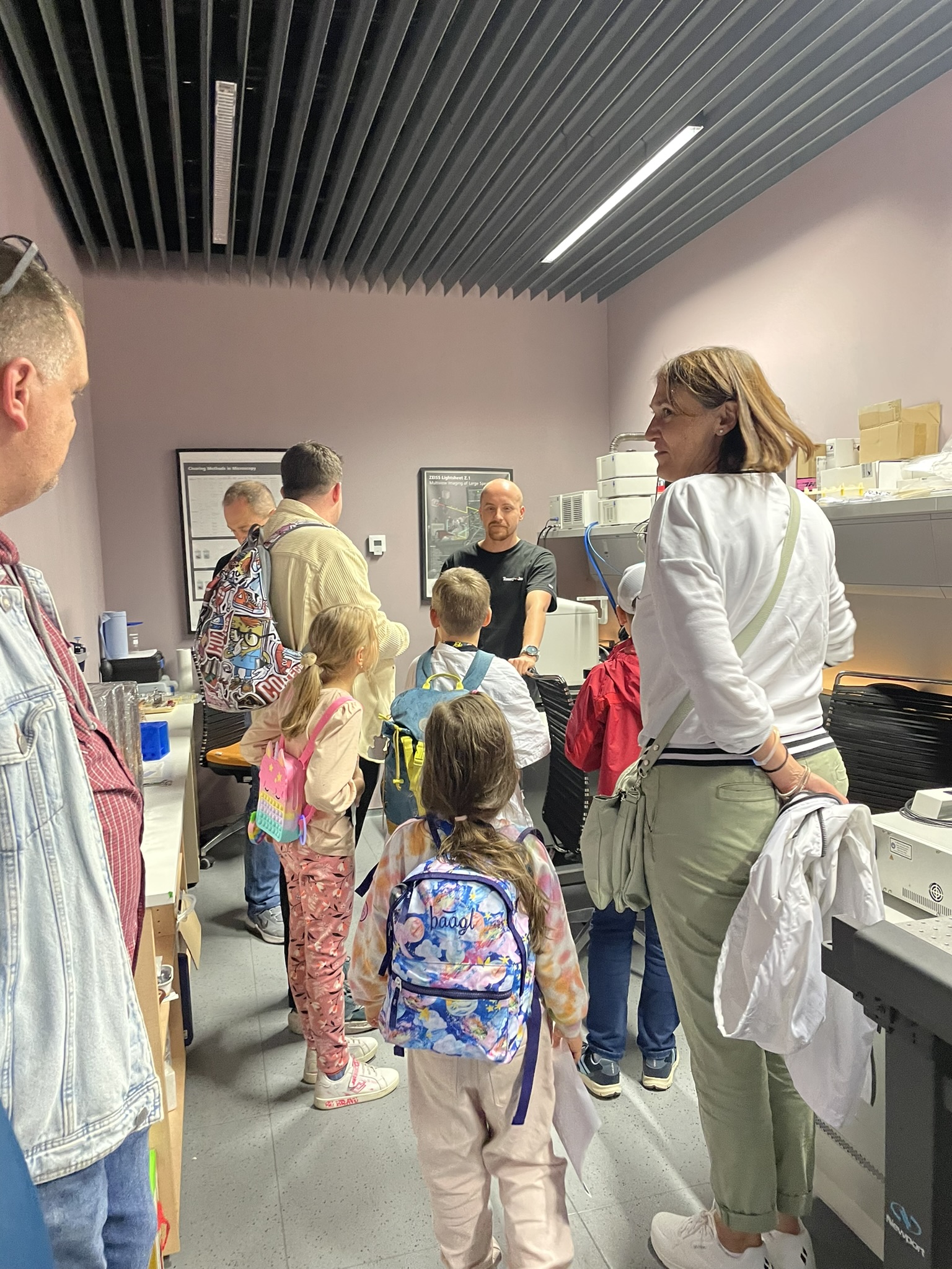
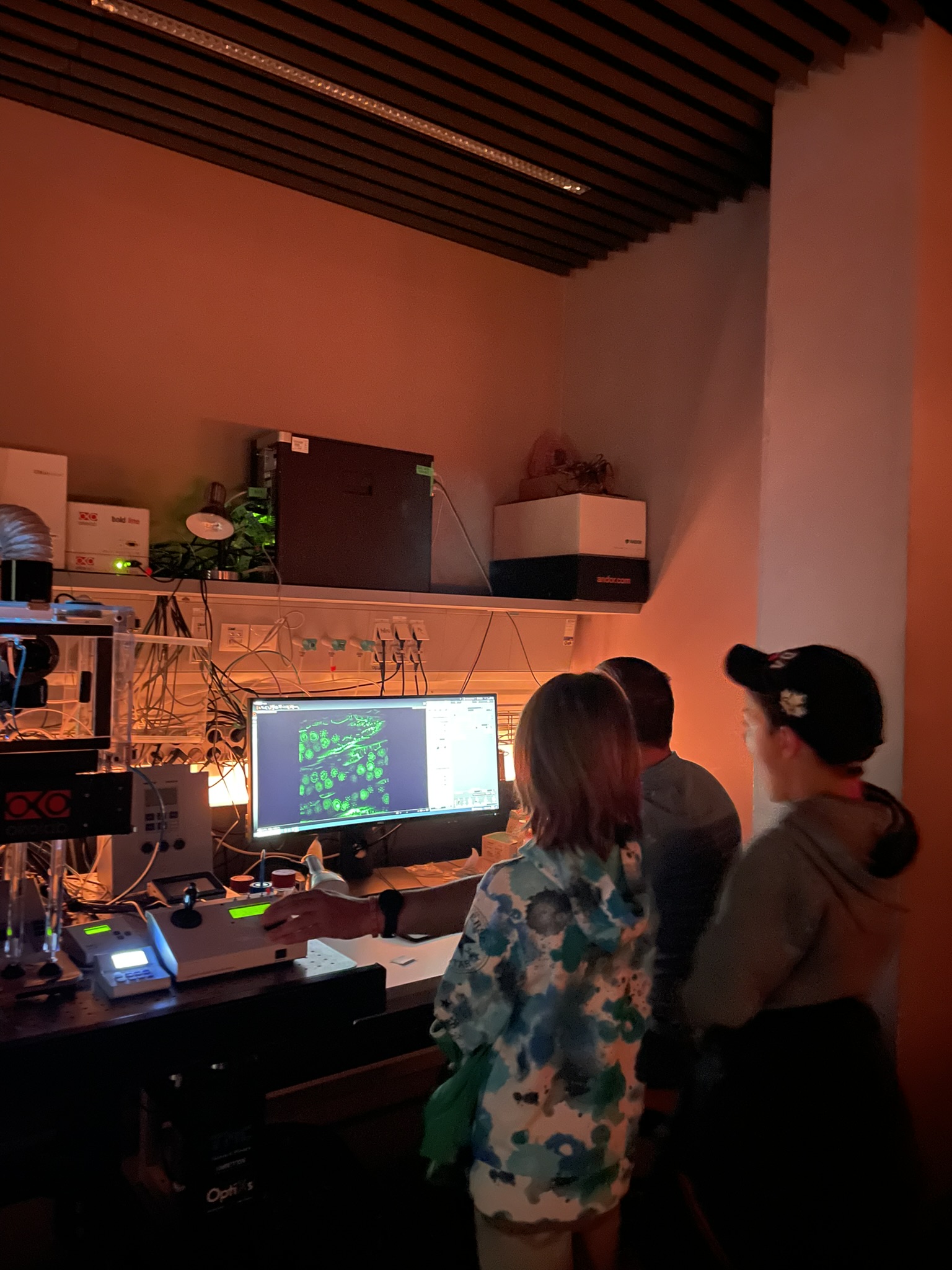
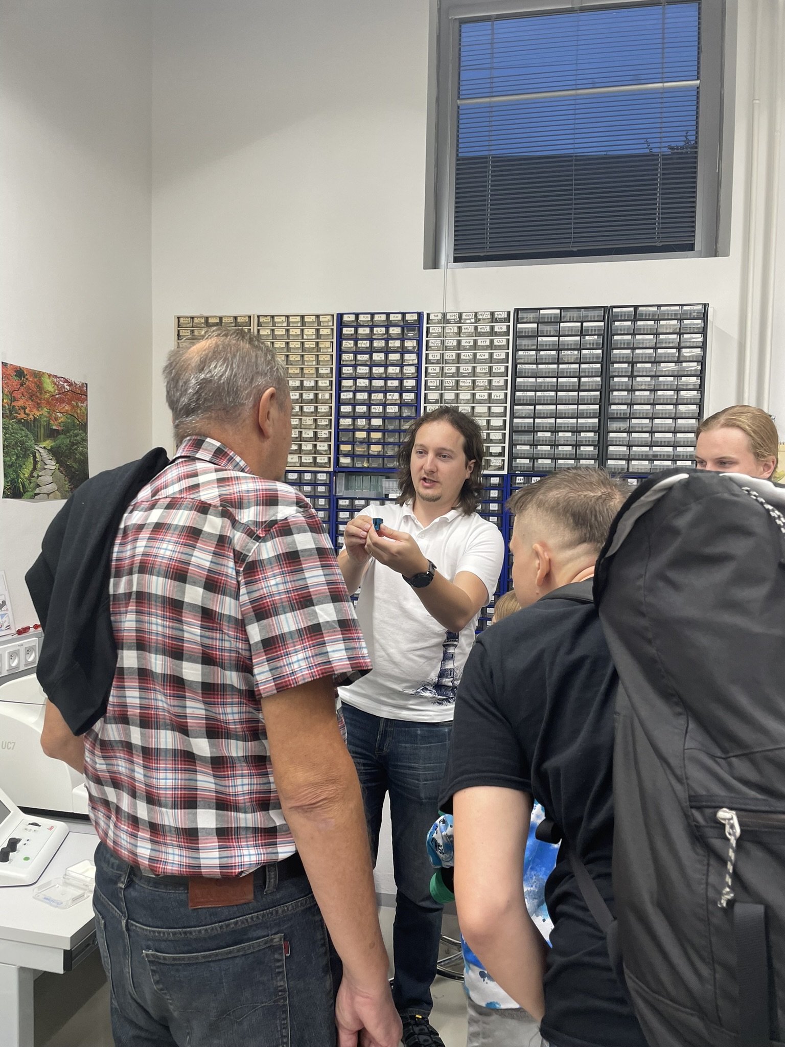
we are thrilled to announce that multiple Czech-BioImaging Core Facilities played a pivotal role in this year’s Czech European Researchers’ Night event, held on September 27th, 2024.
This popular science event, aimed at engaging the general public with the wonders of scientific discovery, saw our microscopy experts showcasing the fascinating world of light microscopy, electron microscopy, medical imaging and digital images produced.
Visitors of all ages had the opportunity to explore the invisible realms through hands-on demonstrations and interactive exhibits at the Institute of Molecular Genetics, Institute of Experimental Medicine, Institute of Physiology, Biology Centre, and Masaryk University – CEITEC. Our team was on hand to explain the latest advancements in microscopy, image analysis and medical imaging how these technologies are revolutionizing research in various fields.
We are proud to contribute to the popularization of science and to inspire the next generation of scientists. Thank you to our partner institutions, who joined this very important event, to the Core Facility staff, who came up with the amazing and very interactove program and everyone who visited our exhibits and made the event a resounding success!
For more information about the event, visit Researchers’ Night: https://www.nocvedcu.cz/en

Dear Czech-BioImaging users,
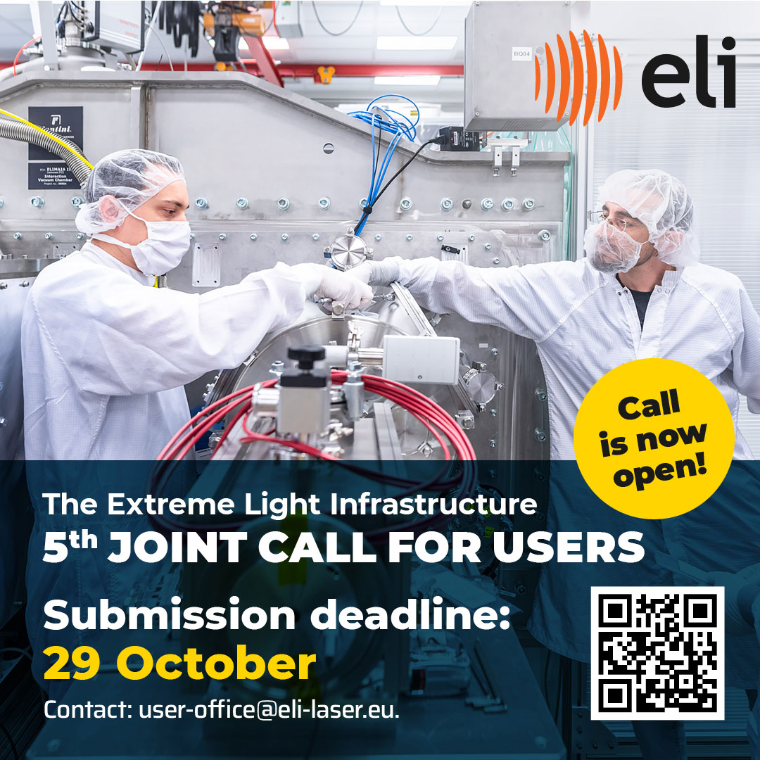
we would like to draw your attention to the 5th ELI User Call with submissions open until 29 October 2024.
Researchers and scientists worldwide are invited to submit their proposals which will be evaluated based on scientific excellence by an international peer-review panel. This Call continues to support a wide range of experiments, leveraging the cutting-edge equipment of:
The complete scientific offer is available on the ELI User Portal, including information about the application and review process.
Proposers are strongly encouraged to contact the technical instrument contacts listed or the ELI User Office [user-office[@]eli-laser.eu].
For more detailed information visit: https://up.eli-laser.eu