27 – 28 May 2025
Institute of Experimental Botany, Czech Academy of Sciences, Rozvojová 263, 165 02 Prague 6
TBD


14 March 2025, 13:00-18:00
Kamenice 5, CEITEC MU, building E35, Brno
Dear Colleagues and Friends,
we are pleased to invite you to the Open Day at the MAFIL CEITEC Core Facility, which is taking place this year as part of the Brain Awareness Week (Týden Mozku) organized by the Czech Academy of Sciences. The Czech-BioImaging staff and users will be available from 1:00 PM to 6:00 PM to share insights into the projects they are currently working on and to discuss any topics related to their scientific focus.
Týden mozku (Week of the Brain) is a unique cycle of lectures on the newest discoveries and trends in brain research and neuroscience, which is a part of the Brain Awareness Week (BAW), the world’s largest public awareness campaign on the success and benefits of brain research. The Week of the Brain in the Czech Republic was initiated and established by the Czech neuroscientist Professor Josef Syka in 1998.
Leading Czech experts in the field of theoretical and clinical neuroscience give lectures during the week-long event, which usually takes place in March. Admission is free to all the festival events. Due to capacity, registration is necessary. We look forward to seeing you there!
For more information (in Czech only), you can visit the official website: www.tydenmozku.cz.

7-11 April 2025
Online
Dear Czech-BioImaging users,
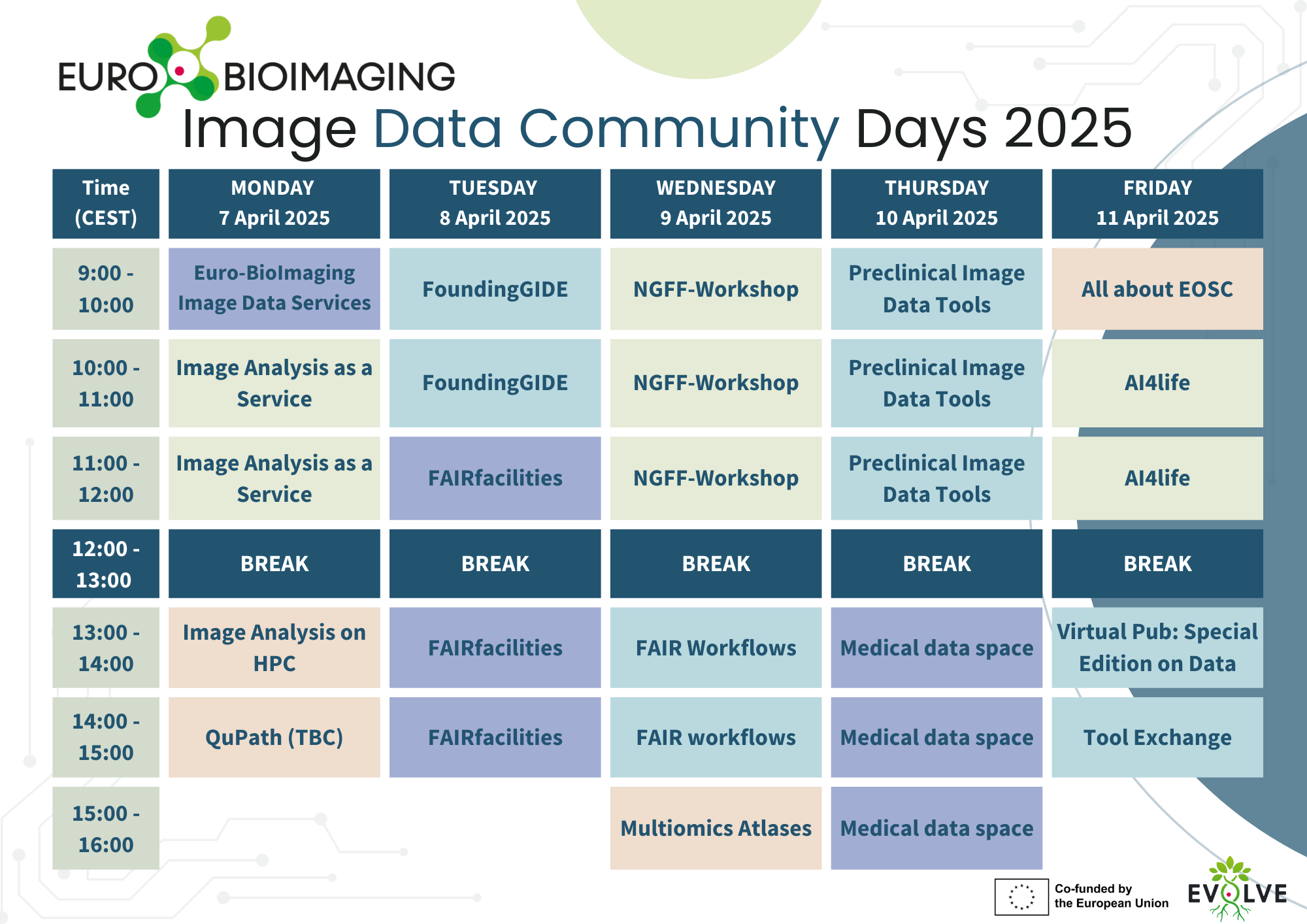
we would like to inform you about the Image Data Community Days that will be happening from 7-11. April this year, organized by Euro-BioImaging.
This free week-long online event will bring together Euro-BioImaging Node staff, researchers and other experts from around the world to explore and exchange on the latest advances in image data through a mix of workshops, lectures and interactive discussions.
Throughout the week, you will have the opportunity to participate in a variety of sessions, ranging from overview talks to in-depth workshops on key topics such as FAIR data, preclinical and medical image data, image analysis, next-generation file formats and AI.
All events will be hosted on Zoom, with sessions scheduled between 9:00 AM to 4:00 PM CEST each day. Register now to secure your spot for this exciting week of learning, collaboration, and networking in the world of image data.
We are very much looking forward to your participation in the week!
Where: Institute of Entomology, Biology Centre of the Czech Acad. Sci., České Budějovice
Dear Czech-BioImaging users,
are you passionate about microscopy and data analysis? The Laboratory of Microscopy and Histology at the Biological Center of the Academy of Sciences of the Czech Republic is seeking a Scientific Assistant to specialize in the analysis of image data from light microscopes. Join us in advancing scientific research and innovation in the field of microscopy!
If interested, please contact via email to sehadova@yahoo.com

13 – 14 October 2025
+ 20 – 21 October 2025
Imaging Methods Core Facility, BIOCEV, Průmyslová 595, 252 50, Vestec
This course is structured in two parts and applicants may register for either part or both:
Introductory Session (October 13-14): Tailored for absolute beginners in Python programming, with a particular emphasis on image analysis and processing. While the skills learned will have broad applications, the focus will center on essential techniques for working with digital images. Prior knowledge of digital imaging is helpful but not required, as all methods will be clearly demonstrated and explained to build a strong foundation in the basics.
Advanced Session (October 20-21): A continuation of the introductory session, focusing on more complex methods, alternative approaches, and hands-on practice with tools like Napari for advanced visualization and AI tools like Cellpose for segmentation. This session builds on skills from the first part, making it ideal for participants who have completed the beginner session and wish to deepen their expertise.
Applicants may register for either part or both.
Covered topics:
09 – 10 December 2025
Building Da I. IPHYS BIF, Krc Campus, Videnska 1083, Prague
The two-day course consists of lectures and hands-on sessions which will demonstrate basic mechanical testing methods such as atomic force microscopy, indentation test or uniaxial and biaxial tensile test. The tests will be introduced in practical sessions together with their biological and methodological relevance. To relate the mechanical tests to the most significant biomechanical structures such as collagen fibers, elastic fibers or fat, the participants will be taught how to link these mechanical properties and microscopy images. Due to the minimum sample alternations, the images are acquired using label-free techniques, for instance, pSHG, THG or CARS.

18 – 21 November 2025
Imaging Methods Core Facility, BIOCEV, Průmyslová 595, 252 50, Vestec
Practical hands-on training on 3D correlative light and electron microscopy combining confocal fluorescence imaging and electron microscopy imaging contain focused ion beam milling (FIB-SEM) and electron tomography (ET).
Light, particularly fluorescence, microscopy offers live-cell compatibility and flexibility in labelling by specific molecular probes. In this way, insight into the function of cells at their native state can be obtained. Electron microscopy, on the hand, provides unmatched spatial resolution. Correlative light and electron microscopy (CLEM) combines the unique information from both techniques in a single image. During the course you will try hands-on a complete CLEM workflow from sample preparation to image processing. The hands-on sessions will be conducted using the state-of-the-art equipment available at IMCF, including high-end fluorescence confocal microscopes, scanning electron microscope (SEM) with focused ion beam (FIB) milling and transmission electron microscope (TEM).
14 – 15 October 2025
Institute of Experimental Botany, Czech Academy of Sciences, Rozvojová 263, 165 02 Prague 6
We are pleased to invite you to the annual two-day course ‘Multimodal Light Microscopy Imaging in Plant Research’ organised by the Imaging Facility IEB in Prague, on 14–15 October 2025.
This year, expect a whirlwind of new technologies presented in four parallel hands-on sessions. Look forward to 3D lifetime STED super-resolution microscopy (Mirava from Abberior Instruments); Lattice Lightsheet 7 (Zeiss); Microfluidics on Vertical stage spinning disk; and Image Analysis and Data Presentation.
The invited speakers are Stefanie Weidkamp-Peters (HHU Duesseldorf); Aleš Benda (Biocev Prague); Gero Schötel (Abberior Instruments); and Pavel Krist (Zeiss).
Tailored to experienced users.
Please register by sending an email to ifieb@ueb.cas.cz. The number of participants is limited to 28.
Registration fee – 1000 CZK.
13 – 17 October 2025
Institute of Molecular Genetics, Vídeňská 1083, 142 20, Prague
The five-day theoretical course with practical demonstrations is devoted to modern light and electron microscopy, and the program is updated every year to reflect the newest trends. The course covers the theoretical background of microscopy as well as fundamental techniques in microscopy, and continues quickly to cutting-edge methods like super-resolution light microscopy, light-sheet microscopy, imaging of whole living organisms, and cryo-electron microscopy.
In the theoretical part, the course will cover the principles of light and electron microscopy, image creation, biological sample preparation recommendations, and will provide a guidance through the principal image processing procedures. The practical part of the course consists of demonstrations in smaller groups at the instruments, covering a wide range of microscopy methods, including Koehler illumination adjustment, widefield fluorescence microscopy, fluorescence macroscopy, confocal scanning and spinning disc microscopy, quantitative phase microscopy, light-sheet microscopy, advanced superresolution microscopy (STED, SIM), transmission electron microscopy, scanning electron microscopy, and cryo-methods of EM sample preparation.
After completing the course, the participant will be able to determine which microscopic technique should be used in order to answer the research question, including the sample preparation and data processing for publication. However, do not expect a detailed technical training on the microscopes or advanced training on sample preparation, super-resolution techniques or electron microscopy, these are part of the more specialized courses which we highly recommend (see course.img.cas.cz for details).
The course is primarily intended for PhD students and young researchers in the biomedical fields. A number of doctoral committees counts this course towards the fulfillment of student’s study obligations.
The course is an introduction to both light and electron microscopy with solid theoretical background extended with many practical presentations. The lectures and also practical sessions are taught by experts and scientists from the field or by product specialists from leading microscopy companies.

November 2025
Viničná Microscopy Core Facility, Viničná 1594/7, 128 00 Prague
TBD
23 – 27 June 2025
Laboratory of Electron Microscopy, Biology Centre of the Czech Academy of Sciences, Branišovská 1160/31 370 05, České Budějovice
TBD
13 May – 14 May 2025
Light Microscopy Core Facility, Institute of Molecular Genetics, Vídeňská 1083, 142 20, Prague
This one-day course focuses intensively on image data segmentation and cell tracking using state-of-the-art deep learning methods like StarDist, Cellpose, Omnipose, and MitoSegNet. It demonstrates how segmentation aids in analyzing image-based objects and how tracking applies segmentation to study cells dynamically over time. TrackMate, a plugin in Fiji, integrates StarDist and Cellpose for cell segmentation and tracking. The course also covers the Delta2 framework for tracking dense bacteria populations and explores ZeroCostDL4Mic, a cloud computing framework with advanced deep learning methods for microscopy tasks like segmentation, object detection, and denoising. The course aims to highlight the practicality and user-friendly nature of these deep learning techniques.
Also, a sponsor’s lecture on Apeer.com, Zeiss’s cloud platform, will focus on image processing, including machine and deep learning. At the course’s end, participants can actively practice segmentation and cell measurement using a virtual reality system from the same company.