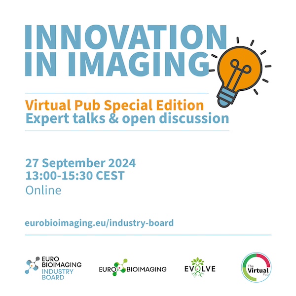
27 September 2024
Online
Dear Czech-BioImaging users,
our colleague, Dominik Pinkas, from the Electron Microscopy Core Facility, Institute of Molecular Genetics of the Czech Academy of Sciences is one of the speakers in the upcoming special edition of the Euro-BioImaging’s Virtual pub on Friday, September 27, 2024, starting at 13:00 CEST.
Come support him and learn how to best exploit your innovation, make it available for others in your facility or partner up with a company.
About the Special edition: https://www.eurobioimaging.eu/events/special-edition-virtual-pub-innovation/
About Virtual pub: https://www.eurobioimaging.eu/our-events/virtual-pub/
Dear Czech-BioImaging users,
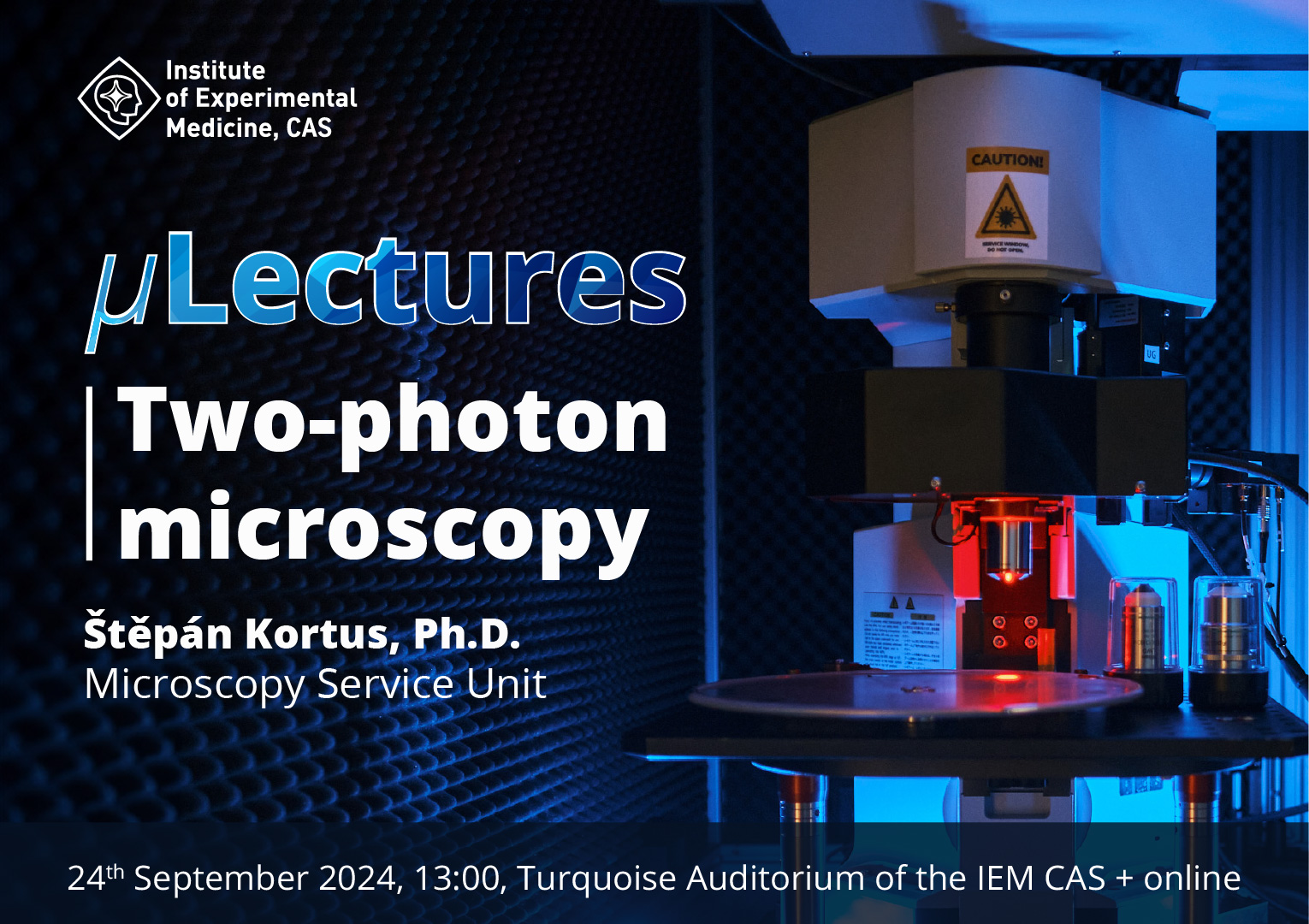
after a summer break, you are invited to the seminar series μLectures organized by the Microscopy Service Centre at the Institute of Experimental Medicine of the Czech Academy of Sciences.
μLectures is designed to enhance researchers’ understanding of microscopy’s fundamental workings and principles. Each session, lasting 30 minutes, focuses on a specific concept or topic within microscopy. The practical information presented aims to deepen researchers’ comprehension of their microscopy experiments, giving them a better overview of the options available and enabling them to improve their experimental design and more effectively identify and resolve potential issues.
The following session is on Two-photon microscopy. This technique is especially suitable for the imaging of processes in vivo. We will explain the two-photon microscopy principle, look at the advantages and disadvantages of this technique and what experiments it can be used for.
When: Tuesday 24. 9. 2024 from 1 p.m.
Where: Turquoise Auditorium of the IEM CAS or online using this link (opens in new window).
Czech-BioImaging supports a newly-launched European Commission economic study to assess the current state of competition in the EU.
As part of this project, the Commission has commissioned an external study to a consortium of firms of which we are part (together with Lear, E.CA Economics, Fideres, Prometeia, and the University of East Anglia).
A part of this study is an external quantitative survey being conducted together with Kantar Group marketing agency. This multi-country survey is carried out among businesses in Europe’s top export sectors and as potential respondents, industrial partners may get contacted to complete the survey for this study.
All information companies provide in the survey will be handled with strict confidentiality and used only for the sole purpose of this study.
See the document from the European Commission outlining the details of the study.
Best regards
CzBI Hub
We would like to inform you of the results of the Elections held in July 2023.
Congratulations to Prof. Julius Lukeš, who was unanimously elected as the Chairman of the Czech-BioImaging Supervisory Board for the period 2023 – 2026 and Prof. Pavel Hozák, proposed by the Coordinator, who was also unanimously elected to continue as Director.
We are very much looking forward to further co-operation.
CzBI Hub
Specion, s.r.o. would like to invite you to a webinar and demonstration of SUPR-DSF (Differential Scanning Fluorimetry) in September at Biocev and Refeyn Mass Photometry in October at IOCB. I will also be happy to share it with relevant groups.
SUPR-DSF (Biocev)
Demo: 9/19/2023
Online Webinar: 8/23/2023
To register for the webinar or demo, use the link below:
https://forms.gle/ZTDfjJzbtUTHqqyr6
Refeyn Mass Photometry (IOCB)
Demo: 9.10. – 10/10/2023
Online Webinar: 10/2/2023
To register for the webinar or demo, use the link below:
https://forms.gle/NcUmyVJEVxikzhiM9
30 June – 4 July 2025
Viničná Microscopy Core Facility, Viničná 1594/7, 128 00 Prague
TBD

The summer school is focused on the advanced methods of biomedical image analysis with emphasis on bio- (microscopy) image analysis. The main goal is to make the students familiar with the advanced state-of-the-art techniques of mathematical morphology, image segmentation, object detection and tracking, synthetic image generation, benchmarking, etc. The focus will be on the methodology suitable for multiple applications.
Special attention will be paid to machine learning approaches. Target groups are Master and Ph.D. students of computer science, mathematics or engineering backgrounds specialized in signal and image processing. Students are supposed to know the basics of signal and image processing, machine learning and have some programming experience in this field.
The main activities of the course will be theoretical lectures supplemented by practical examples run on students’ computers.
The course is focused on theoretical explanation and practical demonstration of advanced methods of HR SEM, such as FIB-SEM, SBF SEM, micro-array tomography, low-voltage STEM and cryo application in biology. The program will also include specimen preparation, basics of 3D reconstruction, and data interpretation.
Single molecule microscopy and manipulation practical course focuses on optical based methods of detection, characterization and even manipulation of single molecules. The aim is to highlight the uniqueness of single molecule based methods for uncovering the mechanisms of molecular interactions in biophysics, but also to show participants the real laboratory world of single molecule experiments. The course covers traditional methods of single molecule observation using TIRF based wide-field microscopy and confocal microscopy combined with spectral and excited state lifetime resolved detection. The single molecule manipulation, including force measurement and confocal imaging of the manipulated molecules, is performed using the optical tweezers. The methodologies covered include single particle tracking and various modifications of fluorescence correlation spectroscopy Single molecule microscopy and manipulation practical course focuses on optical based methods of detection, characterization and even manipulation of single molecules. The aim is to highlight the uniqueness of single molecule based methods for uncovering the mechanisms of molecular interactions in biophysics, but also to show participants the real laboratory world of single molecule experiments. The course covers traditional methods of single molecule observation using TIRF based wide-field microscopy and confocal microscopy combined with spectral and excited state lifetime resolved detection. The single molecule manipulation, including force measurement and confocal imaging of the manipulated molecules, is performed using the optical tweezers. The methodologies covered include single particle tracking and various modifications of fluorescence correlation spectroscopy.
The list of microscopy techniques covered by SMMM course:
Practical workshop/hackathon to learn and improve FIJI open source plugins ImgLib2 and BigDataViewer for analysis/visualization of large datasets, mostly used for lightsheet data processing.
The hackathon is intended for both experienced developers and newcomers, and will include a learnathon part during the first days. Participants are free to work on any topic related to ImgLib2 and/or BigDataViewer, be it improving the core libraries, or using them in other projects. We would try to converge on a few “main” topics beforehand to increase synergies. If you have any particular cool topic ideas, burning needs that could be potentially addressed, etc, please note them in the registration form.
Come and join us in Brno!
The course is an introduction to both light and electron microscopy with solid theoretical background extended with many practical presentations. The lectures and also practical sessions are taught by experts and scientists from the field and also by product specialists from leading microscopy companies.
The five-day theoretical course with practical demonstrations and exercises, is intensively devoted to modern methodologies of light and electron microscopy. Compared to previous years, the program of the course has been updated to copy new trends in microscopy such as super-resolution light microscopy (SIM / PALM, STED, STORM) or Atomic Force Microscopy (AFM). During the course, participants will see practical demonstration of confocal, 2-photon and also cutting-edge superresolution microscopy – STED and SIM. The course also deals with the processing of the image data, however, priority is to acquire practical skills in microscopy techniques (image analysis is the main topic of the course Processing and analysis of microscopic images in biomedicine ).
After completing the course, the participant will be able to determine what is appropriate microscopic technique used to answer the research questions, including the preparation and data processing for publication. Detailed methodological guidance and technical training are part of the more specialized courses.
The course is primarily intended for PhD students and young researchers in the biomedical fields. A number of doctoral committees counts this course towards the fulfillment of student's study obligations. The course will be taught in English.
Imaging in plant research – lectures and hands on
Next Page »« Previous Page