20 – 24 April 2026
First Medical Faculty, Charles University, CAPI, Salmovská 3, 120 00 Prague
Language: Czech
Course for acquiring qualification and professional competence (designing experiments and experimental projects) in the field of experimental animal use according of Act No. 246/1992 On the Protection of Animals Against Cruelty.
Link coming soon
9 – 13 March 2026
First Medical Faculty, Charles University, CAPI, Salmovská 3, 120 00 Prague
Language: English
Course for acquiring qualification and professional competence (designing experiments and experimental projects) in the field of experimental animal use according of Act No. 246/1992 On the Protection of Animals Against Cruelty.
Link coming soon
6-12 September 2026
Centre for Biomedical Image Analysis Faculty of Informatics, Masaryk University, Botanická 68a Brno, 602 00
The summer school is focused on the advanced methods of biomedical image analysis with emphasis on bio- (microscopy) image analysis. The main goal is to make the students familiar with the advanced state-of-the-art techniques of mathematical morphology, image segmentation, object detection and tracking, synthetic image generation, benchmarking, etc. The focus will be on the methodology suitable for multiple applications. Special attention will be paid to the combination of machine learning and classical approaches.
Target groups are Master and Ph.D. students of computer science, mathematics or engineering backgrounds specialized in signal and image processing. Students are supposed to know the basics of signal and image processing, machine learning and have some programming experience in this field. The main activities of the course will be theoretical lectures supplemented by practical examples run on students’ computers.
12-13 + 16-17 February 2026
Imaging Methods Core Facility, BIOCEV, Průmyslová 595, 252 50, Vestec
While the skills learned during the course will have broad applications (data analysis, plotting, scripting…), the focus of exercises will be on image analysis and processing topics. Prior knowledge of digital imaging is helpful but not required.
This course is structured in two parts:
Introductory Session: Tailored for absolute beginners in Python programming. Participants will start with Python installation and basic syntax, progressing to practical exercises focused on working with digital images and relevant libraries for I/O, measurements, ploting and more. Batch processing of multiple images and an intro to Napari (a Python-based interactive image visualization tool) will also be covered.
Advanced Session: This session offers extended hands-on practice, more complex image analysis cases, alternative approaches, and the use of AI tools such as Cellpose for segmentation. This session builds on python skills from the first session. It is ideal for those who completed the introductory session, but also open to participants with basic Python experience. The ability to set up and run a working Python environment is a prerequisite.
Applicants may register for either part or both.
10 – 13 November 2026
Imaging Methods Core Facility, BIOCEV, Průmyslová 595, 252 50, Vestec
Hands-on training on selected time-resolved experimental techniques and data analysis, commonly called Fluorescence Lifetime Imaging Microscopy.
The course focuses on explaining the principles of nano-second time-resolved fluorescence detection, on demonstration of different hardware realizations, on trying various applications of FLIM in biological imaging and on testing of several ways of FLIM data analysis.The aim is to uncover the richness of information hidden in multiparametric fluorescence imaging and to inspire the participants to use the easily obtainable extra contrast in their imaging applications. An important part is to make the FLIM data analysis understandable.
Testing of your own samples is available anytime after (or even before) the FNOB course upon agreement with IMCF staff (may be a subject to instrument fees).
8 – 10 June 2026
Institute of Molecular Genetics, Vídeňská 1083, 142 20, Prague
The three-day course is designed as an intensive hands-on training focused on modern super-resolution microscopy methods and their practical application in biological research. Participants will work directly with advanced imaging systems and create real datasets, gaining practical experience with several super-resolution approaches.
The program combines introductory lectures with extended practical sessions in small groups. The practical part will focus on SIM, SMLM, STED, and optical reassignment approaches, together with data processing and reconstruction workflows. Participants will also learn how to evaluate the suitability of different super-resolution techniques for specific biological questions.
In addition to the technical training, the course will include scientific lectures presenting real biological applications of super-resolution microscopy, providing context for the methods demonstrated during the practical sessions.
The course is open to an international audience of PhD students, postdoctoral researchers, and imaging facility staff interested in advanced light microscopy methods.

12 – 15 October 2026
Imaging Methods Core Facility, BIOCEV, Průmyslová 595, 252 50, Vestec
This course provides an insight into the complexity and principles of sample preparation necessary for ultrastructural analysis by Transmission and Scanning electron microscopy. An emphasis is given to definition of critical steps in sample preparation workflows (from harvesting and fixation to contrasting, immunolabelling, resin embedding, ultrathin sectioning etc.) and their optimization to suit specific type of samples (from nanoparticles to cells, tissues and bio-composites) and to specific aims of the analysis. The practical part of the course will focus primarily on methods suitable for routine quality control and quantitative analysis of particulate and suspension samples including nanoparticles, biopolymers, viruses & bacteria, liposomes, exosomes etc. The EM-SPS course is organized by the Imaging Methods Core Facility at BIOCEV as an annual course supported by the National Infrastructure for Biological and Medical Imaging (Czech-BioImaging, Ministry of Education, Youth and Sports – Large Research Infrastructure, LM2023050).
16 – 17 March 2026
Building Da I. IPHYS BIF, Krc Campus, Videnska 1083, Prague
Course fee is 40 Euros
The two-day course consists of lectures and hands-on sessions which will demonstrate basics in design of experiments, for instance hypothesizing, sampling, data dependency, statistical power or hypothesis testing in biology. The examples will be provided in the field of light microscopy and omics. The participants will learn the ways to correctly acquire data using high-end microscopes using a wide pallet of methods such as FLIM, SHG or Brillouin microscopy or basics of omics approaches. In addition to that the participants will process and analyse the data using traditional approaches, stereology or AI tools in Fiji or Python. Finally, the participants will be taught the data interpretation and presentation.
Although the course is biology science oriented, the skills you will acquire are valid and necessary in any area of research, development or industry.
3 – 5 November 2026
Institute of Molecular Genetics, Vídeňská 1083, 142 20, Prague
The selective practical course “Live cell imaging” is an intensive educational block focused on the investigation of living cells and whole organisms using light and fluorescence microscopy. During the course, 12-15 participants will learn about a wide range of instruments using a variety of methods suitable for imaging live specimens. They will learn what methods can be chosen for a given type of specimen, what their advantages are, as well as what their limitations and shortcomings are. They will also learn about the possibilities of studying the kinetics of processes in living cells and the procedures for preparing samples containing living cells, tissues and organisms.
The morning of each of the three days is filled with lectures on microscopy systems and methods, including several short scientific papers aimed at demonstrating the data that can be obtained by these approaches. The emphasis is then primarily on the afternoon practical part of the course, where participants will try their hand at working with a selection of microscopes on real live specimens. During the hands-on sessions, participants will be motivated to actively participate in the set-up of each instrument, data acquisition and subsequent data evaluation. Day one will focus primarily on widefield systems, day two will focus on confocal methods, and day three will cover advanced methods including superresolution and kinetic measurements.
Refreshments and meals are provided throughout the course.
26 – 27 May 2026
Institute of Experimental Botany, Czech Academy of Sciences, Rozvojová 263, 165 02 Prague 6
We are pleased to invite you to the annual two-day course Basics of Light Microscopy in Plant Research organised by the Imaging Facility IEB in Prague.
This time, we’re going back to the fundamentals, to the very roots. A solid understanding of basic fluorescence light microscopy is essential for all of us, and this course is designed to strengthen it.
Our hands-on sessions will focus on practical, everyday techniques, including:
As always, we will also cover bioimage analysis, along with applied scientific lectures.
If you are a pre-graduate student, PhD student, or researcher, don’t hesitate to register—this course is for you.
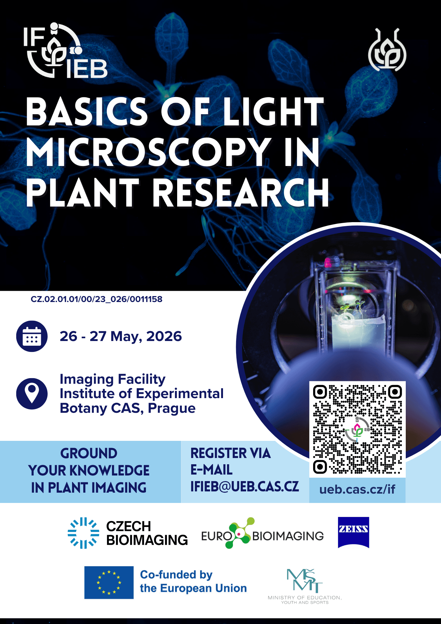
Register via e-mail ifieb@ueb.cas.cz

13 – 14 October 2026
Institute of Experimental Botany, Czech Academy of Sciences, Rozvojová 263, 165 02 Prague 6
We are pleased to invite you to the annual two-day course Advanced Multimodal Light Microscopy Imaging in Plant Research’ organised by the Imaging Facility IEB in Prague.
More TBA
TBA
19 – 21 May 2026
Light Microscopy Core Facility, Institute of Molecular Genetics, Vídeňská 1083, 142 20, Prague
This one-day course focuses intensively on image data segmentation and cell tracking using state-of-the-art deep learning methods like StarDist, Cellpose, Omnipose, and MitoSegNet. It demonstrates how segmentation aids in analyzing image-based objects and how tracking applies segmentation to study cells dynamically over time. TrackMate, a plugin in Fiji, integrates StarDist and Cellpose for cell segmentation and tracking. The course also covers the Delta2 framework for tracking dense bacteria populations and explores ZeroCostDL4Mic, a cloud computing framework with advanced deep learning methods for microscopy tasks like segmentation, object detection, and denoising. The course aims to highlight the practicality and user-friendly nature of these deep learning techniques.
Also, a sponsor’s lecture on Apeer.com, Zeiss’s cloud platform, will focus on image processing, including machine and deep learning. At the course’s end, participants can actively practice segmentation and cell measurement using a virtual reality system from the same company.

