25 June 2026
Institute of Molecular Genetics, Vídeňská 1083, 142 20, Prague
Dear Users,
we warmly invite you to join us for an afternoon and evening in an unusual setting, away from the dark microscopy rooms. The event will offer discussions not only about microscopy, but also entertainment, good food, and refreshing drinks. Together, we will celebrate another successful year of collaboration and perhaps recharge for the year ahead. The event is organized by the Prague microscopy facilities united within the Czech-BioImaging infrastructure and the Prague Node of Euro-BioImaging ERIC and hosted by the Institute of Molecular Genetics of the Czech Academy of Sciences (IMG).
We kindly ask everyone planning to attend to register in advance. This will help us estimate the required amount of food and drinks. After registering, you will receive a confirmation e-mail, which will also serve as your entry pass to the Krč campus at the main gate.
Deadline: 18 June 2026
Looking forward to seeing you!
Organizing Team


1-4 December 2026
To Be Announced
This comprehensive course is designed specifically for biologists to master cutting-edge image analysis software. It covers both the theory of image processing and its practical applications, focusing on precise and efficient image quantification, segmentation, and visualization techniques. Participants will gain hands-on experience with industry-leading commercial and open-source tools and elevate their data analysis skills to the next level.
The course also introduces the FAIR principles for scientific data and demonstrates effective strategies for data management using the OMERO database.
The course is organised by the Cellular Imaging Core Facility in collaboration with the Faculty of Informatics, Masaryk University.
8-11 September 2026
CEITEC MU, University Campus, Building C02, CELLIM CF, Brno
The Advanced Light Microscopy Methods is an advanced course for students, postdocs, technicians, and other laboratory staff in biology and biomedical science who require specialised microscopy methods for their experiments. The course consists of theoretical lectures (25%) and practical hands-on sessions on selected microscopes (50%) and image processing software (25%).
Overall, this course provides an overview of core principles behind super-resolution, FLIM-FRET and mesoscopic imaging methods, together with corresponding essential image processing steps. Participants will learn theoretical principles behind large sample imaging, super-resolution methods like Airyscan, Optical Reassignment, Structured Illumination and Single Molecule Localisation, as well as Fluorescence Lifetime and Förster Resonance Energy Transfer Microscopy.
A dedicated practical session will demonstrate the learned principles directly on real microscope acquisitions. Participants will gain skills needed to operate selected microscopes and experience how to setup advanced experimental workflow, and perform the most common troubleshooting.
The workshop concludes with an overview of related workflows in image processing and analysis utilising related software (ZEN, LAS X). Finally, the participants will receive comprehensive guidelines for advanced analysis and image visualisation in Imaris.
Practical training will be conducted on the following systems:
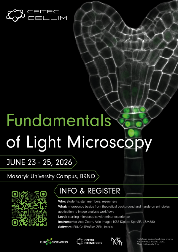
23-25 June 2026
CEITEC MU, University Campus, Building C02, CELLIM CF, Brno
The Fundamentals of Light Microscopy is a basic course for students, postdocs, technicians, and other laboratory staff in biology and biomedical science who require widefield and/or confocal microscopy for their experiments. The course consists of theoretical lectures (50%) and practical hands-on session on selected microscopes and software (50%).
Overall, this course provides an introduction to the fundamentals of light microscopy, with a focus on understanding how images are formed and how acquisition choices affect resolution, contrast, and quantitative image quality. Participants will learn core principles including diffraction and the point spread function, numerical aperture and refractive index effects, sampling and digitization, detector characteristics and noise, and the conceptual differences between major imaging modalities such as widefield, confocal, super-resolution and light sheet microscopy.
A dedicated practical session will demonstrate the learned principles directly on real microscope acquisitions. Participants will gain skills needed to operate selected microscopes and experience how changing parameters influences contrast, optical sectioning, signal-to-noise ratio, and artifacts.
Finally, the workshop concludes with an introduction to basic workflows in image processing and analysis, including foundational steps for viewing, correcting, and preparing microscopy datasets for downstream quantification using open-source and commercial tools (FIJI/ImageJ, CellProfiler, ZEN, Imaris).
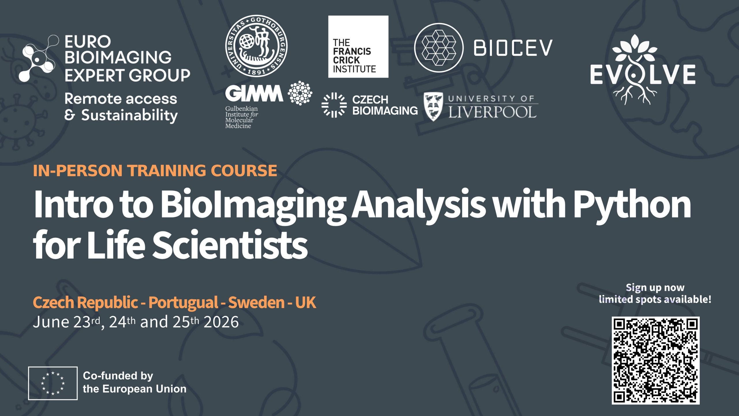
23 – 25 June 2026
Czech Republic, Portugal, Sweden, UK
Průmyslová 595, 252 50 Vestec, Czech Republic
This course is jointly organised by four Euro-BioImaging Nodes and will be run at four connected training centres that are part of Euro-BioImaging:
This course is organised under the EVOLVE project (Grant agreement N° 101130986) as part of Euro-BioImaging’ effort in increasing the adoption of hybrid training thus combining the advantages of remote and in-person training.
This distributed course will be held in person at the four locations and the four local training hubs will be remotely connected and jointly going through the training programme provided each day by one of the sites.
This means that students can enjoy the advantages of small in-person training – connecting with their fellow students and direct support from their trainers – as well as being connected on an international level and benefit from the training and image analysis expertise at all the training centres.
The course is designed for life science researchers with some Image Analysis experience who want to push their analysis capabilities to the next level by using Python and Jupyter Notebooks, as well as implementing and applying analysis tools developed by the community for their image analysis challenges.
Financial support to cover travel and accommodation (Max. 500 EUR per person) is available upon request and for a limited number of participants.
Registration is open until April 15th, and local travel grants are available here.
The BioImaging Facility at the Institute of Physiology of the Czech Academy of Sciences (IPHYS) has recently expanded its technological portfolio with a new Brillouin microscopy system. The acquisition was made possible through the OP JAK project funded by the Ministry of Education, Youth and Sports of the Czech Republic (MEYS) and represents a strategic investment into progressive technologies that were previously not available in open access within Czech-BioImaging or elsewhere in Czechia. As part of the national infrastructure Czech-BioImaging and the Euro-BioImaging Advanced Light and Electron Microscopy Prague Node, the facility now offers Brillouin microscopy in open access to the scientific community.
Šimon Vrana, Imaging Specialist at IPHYS BioImaging Facility supports users in applying advanced biophysical imaging methods to their research questions: “I am specialized in Atomic Force Microscopy, Brillouin and other biophysical methodologies. My role in the facility is to support our users with different imaging approaches to address scientific questions in this field of biomechanics.”

Brillouin microscopy is an emerging optical modality that enables non-contact, label-free mapping of mechanical properties in biological samples. The technique measures the interaction between laser light and thermally driven acoustic waves within a sample, providing information about its internal mechanical characteristics.
By extracting parameters such as the longitudinal elastic modulus and viscoelastic behavior, researchers can investigate how stiffness and mechanical heterogeneity influence biological systems. This is particularly relevant because changes in mechanical properties are often associated with development, differentiation, and disease.
Although Brillouin microscopy has been widely used in cell and tissue research, the first users of the new system at IPHYS are working in plant biology. Current projects include measurements on Physcomitrella moss provided by Imaging Facility Institute of Experimental Botany of the Czech Academy of Sciences, comparing genetically modified lines with wild-type counterparts.
The system is suitable for a broad spectrum of samples:
In general, any sample compatible with confocal microscopy can be investigated, provided it is sufficiently transparent and preferably adherent.
The facility invested in a commercial Brillouin microscope (Discoverer, CellSense) to ensure reliability, robustness, and accessibility for users. Unlike many custom-built systems, the commercial platform enables streamlined operation and faster onboarding.
“We want to offer Brillouin Microscopy as a service, and the CellSense system is very reliable and user-friendly. After a short training and some troubleshooting, users should be able to measure samples themselves.”
This strategic choice allows Czech-BioImaging to provide sustainable open access to technology without the need for extensive in-house development and maintenance.
A key advantage of the IPHYS setup is its integration of Brillouin microscopy with spinning disk fluorescence microscopy, including the Yokogawa SoRa super-resolution system. This multimodal configuration enables researchers to correlate mechanical mapping with high-resolution fluorescence imaging.
In practice, this means that users can:
“Users can examine a cell sample stained with fluorescent dyes, identify a protein of interest, and then shift to Brillouin microscopy to measure local viscoelastic properties without dyes or mechanical probes.”
Beyond Brillouin imaging, the facility provides access to confocal and multiphoton microscopy, FLIM, PLIM, light-sheet imaging, and mechanical testing approaches such as AFM and compression, biaxial tensile testing—allowing comprehensive multimodal experimental design.
The facility primarily supports projects where biomechanics plays a central role in the biological question. Researchers can investigate:
By integrating Brillouin microscopy into broader imaging workflows, scientists can better understand how physical properties shape biological function.
As part of Czech-BioImaging, the IPHYS BioImaging Facility operates under a 100% open-access policy and provides comprehensive user support.
Services include:
While commercial software provides basic Brillouin data processing, the facility also supports advanced workflows using third-party tools such as Fiji and develops customized solutions in C++, C#, or Python when required.
With the introduction of Brillouin microscopy, Czech-BioImaging strengthens its position within Euro-BioImaging ERIC.
The combination of:
makes the IPHYS BioImaging Facility a unique access point for researchers interested in biomechanics-driven biological questions in Czechia and Europe.
21 – 23 April 2026
Institute of Molecular Genetics, Vídeňská 1083, 142 20, Prague
The international conference Vision for Life – Innovation Days in Bioscience Imaging will bring three
days of intensive knowledge exchange, technological innovation, and hands-on experience in
advanced microscopy and biological imaging to Prague.
The event will take place at the Institute of Molecular Genetics of the Czech Academy of Sciences
(IMG). It is organized by Specion (an authorized distributor of Leica Microsystems) in collaboration
with IMG.
The conference is fully supported by the Light Microscopy Core Facility at IMG, which is part of the
Czech BioImaging national research infrastructure and a member of the Prague node of Euro-
BioImaging, the European research infrastructure for biological and biomedical imaging.
Pavel Hozák, Director of Czech BioImaging, will contribute to the program with a presentation titled “Czech-BioImaging – The National Imaging Platform for All,” introducing the national research infrastructure and its mission to provide open access to state-of-the-art imaging technologies.
The program will feature:
The main keynote speakers of this year’s edition include:
The conference will connect the scientific community, technology partners, and core facility
professionals, creating a platform to discuss current challenges and future directions in bioscience
imaging at both the national and European level.
For any questions regarding the organization of the event, please contact Martin Kopecky (Specion),
main organizer.
8 – 13 October 2026
First Medical Faculty, Charles University, CAPI, Salmovská 3, 120 00 Prague
Language: English
The event “Imaging Horizons”, organized by the Center for Advanced Preclinical Imaging (CAPI) at the First Faculty of Medicine, Charles University, aims to raise awareness of modern imaging methods and their importance for preclinical research. The goal is to present the wide range of technologies available at CAPI, including MRI, PET, CT, ultrasound, photoacoustic imaging, in vivo fluorescence, and magnetic particle imaging, and to demonstrate their practical applications in medicine and biomedicine.
The program combines a 2.5-hour block of expert lectures featuring real data from CAPI projects, followed by a networking session to foster collaboration between participants and experts. The lecture block will be streamed online to enable interactive engagement with a broader audience and increase the event’s reach. Another part of the program consists of guided tours of CAPI laboratories, organized in four sessions (two on the day of the lectures and two on a separate day close to the event date). Participants will be divided into small groups and will learn about measurement principles, sample preparation, instrument demonstrations, and typical data outputs.
The event targets advanced master’s students, PhD candidates, and early-career researchers from medicine, biomedicine, biology, physics, chemistry, and related disciplines. A unique feature of CAPI is its multimodal approach, in-house technology development, and integration into the European Euro-BioImaging infrastructure, which facilitates connections with the international scientific community and supports new collaborations.
Link coming soon
2 – 4 December 2026
Institute of Experimental Medicine, Vídeňská 1083, 142 20, Prague
This course is aimed at participants familiar with the basics of image processing, who want to automate their analysis using scripting. We will work with ImageJ macro language and use it to create a workflow for segmentation, batch processing and analysis of fluorescence microscopy images. In the second part of the course, we will introduce Cellpose, a machine learning algorithm for cell segmentation, demonstrate its use through command line and ImageJ plugin, and incorporate its output into the ImageJ workflow.
No previous programming knowledge is expected, and the course will therefore also briefly introduce basic programming concepts, such as variables, for loops and conditionals (if-else statements). Most of the sessions will be hands-on, and the last day will be dedicated to individual consultations of user projects and questions.
The course is primarily aimed at participants with some experience with ImageJ and basic understanding of bioimage analysis. We expect no or minimal knowledge of programming.
Participants are required to bring their own laptop for hands-on exercises. If you do not have access to a laptop, please inform us in advance, and we will provide you with one. Prior to the event, you will receive a list of the required software programs to install for the course.
Please make sure that all required software is installed and works prior to the workshop, especially if your laptop runs macOS or Linux (we work mostly with Windows and are therefore not as familiar with the other operating systems, therefore troubleshooting during the course is difficult).
28 – 29 May 2026
Institute of Experimental Medicine, Vídeňská 1083, 142 20, Prague
This course introduces participants to the fundamentals of volumetric and time-lapse microscopy data processing, with a focus on visualization, segmentation, and quantitative analysis. The first day offers a hands-on introduction to Imaris, covering its core modules (Spots, Surfaces, and Filaments). The second day builds on this and includes a broader discussion on big data challenges, alternative 3D processing tools in ImageJ/Fiji, and options for data storage, sharing, and reuse (FAIR data).
The course is organized in hybrid form and it is possible to join either in-person or online.
Imaris is the main software used, and the course is designed to provide a comprehensive overview of its capabilities. As such, it is primarily aimed at participants with no or limited experience. However, advanced features and exercises will also be included to benefit those already familiar with the software.
24 – 25 November 2026
Building Da I. IPHYS BIF, Krc Campus, Videnska 1083, Prague
Course fee is 40 Euros
The two-day course consists of lectures and hands-on sessions which will demonstrate basic mechanical testing methods such as atomic force microscopy, Brillouin microscopy, indentation test or uniaxial and biaxial tensile test. The tests will be introduced in practical sessions together with their biological and methodological relevance. To relate the mechanical tests to the most significant biomechanical structures such as collagen fibers, elastic fibers or fat, the participants will be taught how to link these mechanical properties and microscopy images. Due to the minimum sample alternations, the images are acquired using label-free techniques, for instance, pSHG, THG or CARS or confocal reflectance microscopy.
1 – 4 December 2026
Institute of Scientific Instruments of the Czech Academy of Sciences, Královopolská 147, Brno, Czech Republic
The three/four-day (TBD) course organized by the ISI EM team at the Institute of Scientific Instruments of the Czech Academy of Sciences will offer students and young researchers in the fields of life sciences and soft materials both theoretical and practical training in electron microscopy and Raman spectroscopy. Participants will gain hands-on experience with SEM and FIB-SEM microscopes, biological sample preparation, EDX analysis, and cryo-SEM workflow. The program will combine introductory lectures with intensive hands-on sessions in small groups and will be complemented by a social evening and discussion sessions at the end of each day.
Link coming soon

