10 – 13 November 2026
Imaging Methods Core Facility, BIOCEV, Průmyslová 595, 252 50, Vestec
Hands-on training on selected time-resolved experimental techniques and data analysis, commonly called Fluorescence Lifetime Imaging Microscopy.
The course focuses on explaining the principles of nano-second time-resolved fluorescence detection, on demonstration of different hardware realizations, on trying various applications of FLIM in biological imaging and on testing of several ways of FLIM data analysis.The aim is to uncover the richness of information hidden in multiparametric fluorescence imaging and to inspire the participants to use the easily obtainable extra contrast in their imaging applications. An important part is to make the FLIM data analysis understandable.
Testing of your own samples is available anytime after (or even before) the FNOB course upon agreement with IMCF staff (may be a subject to instrument fees).
8 – 10 June 2026
Institute of Molecular Genetics, Vídeňská 1083, 142 20, Prague
The three-day course is designed as an intensive hands-on training focused on modern super-resolution microscopy methods and their practical application in biological research. Participants will work directly with advanced imaging systems and create real datasets, gaining practical experience with several super-resolution approaches.
The program combines introductory lectures with extended practical sessions in small groups. The practical part will focus on SIM, SMLM, STED, and optical reassignment approaches, together with data processing and reconstruction workflows. Participants will also learn how to evaluate the suitability of different super-resolution techniques for specific biological questions.
In addition to the technical training, the course will include scientific lectures presenting real biological applications of super-resolution microscopy, providing context for the methods demonstrated during the practical sessions.
The course is open to an international audience of PhD students, postdoctoral researchers, and imaging facility staff interested in advanced light microscopy methods.

13 – 16 April 2026
Imaging Methods Core Facility, BIOCEV, Průmyslová 595, 252 50, Vestec
This course provides participants with an insight into the complexity and principles of biological sample preparation necessary for routine ultrastructural analysis by Transmission or Scanning electron microscopy. An emphasis is given to definition of critical steps in sample preparation workflows and their optimization to suit specific type of samples (from nanoparticles to cells, tissues and bio-composites) and to specific aims of the analysis. The practical part of the course will cover sample harvesting, chemical fixation, contrasting, resin embedding, ultra-thin sectioning, negative staining, critical point drying, plunge freezing and more. The EM-SPS course is organized by the Imaging Methods Core Facility at BIOCEV as an annual course supported by the National Infrastructure for Biological and Medical Imaging (Czech-BioImaging, Ministry of Education, Youth and Sports – Large Research Infrastructure, LM2023050).
16 – 17 March 2026
Building Da I. IPHYS BIF, Krc Campus, Videnska 1083, Prague
Course fee is 40 Euros
The two-day course consists of lectures and hands-on sessions which will demonstrate basics in design of experiments, for instance hypothesizing, sampling, data dependency, statistical power or hypothesis testing in biology. The examples will be provided in the field of light microscopy and omics. The participants will learn the ways to correctly acquire data using high-end microscopes using a wide pallet of methods such as FLIM, SHG or Brillouin microscopy or basics of omics approaches. In addition to that the participants will process and analyse the data using traditional approaches, stereology or AI tools in Fiji or Python. Finally, the participants will be taught the data interpretation and presentation.
Although the course is biology science oriented, the skills you will acquire are valid and necessary in any area of research, development or industry.
3 – 5 November 2026
Institute of Molecular Genetics, Vídeňská 1083, 142 20, Prague
The selective practical course “Live cell imaging” is an intensive educational block focused on the investigation of living cells and whole organisms using light and fluorescence microscopy. During the course, 12-15 participants will learn about a wide range of instruments using a variety of methods suitable for imaging live specimens. They will learn what methods can be chosen for a given type of specimen, what their advantages are, as well as what their limitations and shortcomings are. They will also learn about the possibilities of studying the kinetics of processes in living cells and the procedures for preparing samples containing living cells, tissues and organisms.
The morning of each of the three days is filled with lectures on microscopy systems and methods, including several short scientific papers aimed at demonstrating the data that can be obtained by these approaches. The emphasis is then primarily on the afternoon practical part of the course, where participants will try their hand at working with a selection of microscopes on real live specimens. During the hands-on sessions, participants will be motivated to actively participate in the set-up of each instrument, data acquisition and subsequent data evaluation. Day one will focus primarily on widefield systems, day two will focus on confocal methods, and day three will cover advanced methods including superresolution and kinetic measurements.
Refreshments and meals are provided throughout the course.
26 – 27 May 2026
Institute of Experimental Botany, Czech Academy of Sciences, Rozvojová 263, 165 02 Prague 6
We are pleased to invite you to the annual two-day course Basics of Light Microscopy Imaging in Plant Research organised by the Imaging Facility IEB in Prague.
More TBA
TBA

13 – 14 October 2026
Institute of Experimental Botany, Czech Academy of Sciences, Rozvojová 263, 165 02 Prague 6
We are pleased to invite you to the annual two-day course Advanced Multimodal Light Microscopy Imaging in Plant Research’ organised by the Imaging Facility IEB in Prague.
More TBA
TBA
19 – 21 May 2026
Light Microscopy Core Facility, Institute of Molecular Genetics, Vídeňská 1083, 142 20, Prague
This one-day course focuses intensively on image data segmentation and cell tracking using state-of-the-art deep learning methods like StarDist, Cellpose, Omnipose, and MitoSegNet. It demonstrates how segmentation aids in analyzing image-based objects and how tracking applies segmentation to study cells dynamically over time. TrackMate, a plugin in Fiji, integrates StarDist and Cellpose for cell segmentation and tracking. The course also covers the Delta2 framework for tracking dense bacteria populations and explores ZeroCostDL4Mic, a cloud computing framework with advanced deep learning methods for microscopy tasks like segmentation, object detection, and denoising. The course aims to highlight the practicality and user-friendly nature of these deep learning techniques.
Also, a sponsor’s lecture on Apeer.com, Zeiss’s cloud platform, will focus on image processing, including machine and deep learning. At the course’s end, participants can actively practice segmentation and cell measurement using a virtual reality system from the same company.
13 – 17 April 2026
Light Microscopy Core Facility, Institute of Molecular Genetics, Vídeňská 1083, 142 20, Prague
The course will delve into fundamental aspects of image data acquisition, processing, and analysis, encompassing techniques in stereology. Alongside theoretical principles, the course will prioritize hands-on practical learning. Participants will gain proficiency in utilizing the freely available software package Fiji for both basic and advanced analyses. They will learn to assess co-localizations, analyze data from FRAP and electron microscopy, track particles, segment objects in images, and explore methods of employing artificial intelligence for image segmentation.
Additionally, participants will master techniques to enhance data quality through deconvolution using Huygens software. An interactive session featuring Imaris software will be complemented by practical exercises. Furthermore, this year introduces a new series of practical sessions utilizing Napari, a Python-based environment for image processing. Independent practical tasks involving Fiji and Huygens will also constitute a significant component of the course.
12 – 16 October 2026
Institute of Molecular Genetics, Vídeňská 1083, 142 20, Prague
The course is an introduction to both light and electron microscopy with solid theoretical background extended with many practical presentations. The lectures and also practical sessions are taught by experts and scientists from the field and also by product specialists from leading microscopy companies. The five-day theoretical course with practical demonstrations and exercises, is intensively devoted to modern methodologies of light and electron microscopy. Compared to previous years, the program of the course has been updated to copy new trends in microscopy such as super-resolution light microscopy (SIM / PALM, STED, STORM) or Atomic Force Microscopy (AFM).
During the course, participants will see practical demonstration of confocal, 2-photon and also cutting-edge superresolution microscopy – STED and SIM. The course also deals with the processing of the image data, however, priority is to acquire practical skills in microscopy techniques (image analysis is the main topic of the course Processing and analysis of microscopic images in biomedicine ). After completing the course, the participant will be able to determine what is appropriate microscopic technique used to answer the research questions, including the preparation and data processing for publication.
Detailed methodological guidance and technical training are part of the more specialized courses. The course is primarily intended for PhD students and young researchers in the biomedical fields. A number of doctoral committees counts this course towards the fulfillment of students’ study obligations. The course will be taught in English.
The course is jointly organized with the IPHYS BIF core facility.
17-18 September 2026
CEITEC Brno University of Technology, B0.04, Purkyňova 123, Brno
Digital holographic, interference, fluorescence and confocal microscopy and its use in modern medicine, deployment of live cells, operating the Q-Phase microscope
TBA
Dear Czech-BioImaging Users and Friends,
with the beginning of the new year, we are pleased to announce the launch of the new visual identity of Czech-BioImaging. This update reflects the steady development and growth of our research infrastructure, as well as our effort to keep pace with recent trends in imaging and scientific communication. It also mirrors the expanding user community we serve and our ongoing commitment to quality, scientific excellence, reliability, and continuity.
Our new logo builds on the foundations of the previous one. It retains our signature blue colour to preserve continuity, while its simplified and modernised form improves clarity and readability across formats. The refreshed visual identity introduces a unified system that strengthens our communication across all facilities and enhances how we present Czech-BioImaging to the wider public.
Why we refreshed our identity
As Czech-BioImaging continues to grow, so does our responsibility to communicate clearly and consistently with our users, collaborators, and partners in the Czech Republic and abroad. A strong and coherent visual identity helps us represent who we are today and how our infrastructure has evolved over the years.
The refreshed identity enables us to:
The new identity is the outcome of a thoughtful design process carried out in collaboration with graphic designer Martin Vosátka. It is grounded in our long-term mission, shaped by feedback from our community, and guided by the principles set during the development of our brand.
A logo inspired by imaging
The new Czech-BioImaging logo is grounded in the visual language of imaging.
Its concentric, rotational elements reference lens adjustment and focus, symbolising precision, clarity, and the interconnected network of our facilities.
The accompanying logotype ensures readability and a modern, clean presentation suitable for both digital and print environments.
Together, the symbol and wordmark create a clear and flexible logo system that represents our scientific focus and the collaborative nature of Czech-BioImaging.

A colour palette built for clarity and impact
Our updated colour palette is designed to support clear communication while remaining visually distinctive. It is based on four primary tones:
A complementary secondary palette expands the system for posters, outreach materials, and digital communication, allowing flexibility without losing consistency.
Typography that supports readability
The primary typeface, Open Sauce One, was chosen for its balance of modern design and excellent readability. A clear typographic structure helps us communicate scientific content in a way that is accessible, precise, and visually coherent. Where required, Roboto serves as a reliable replacement.

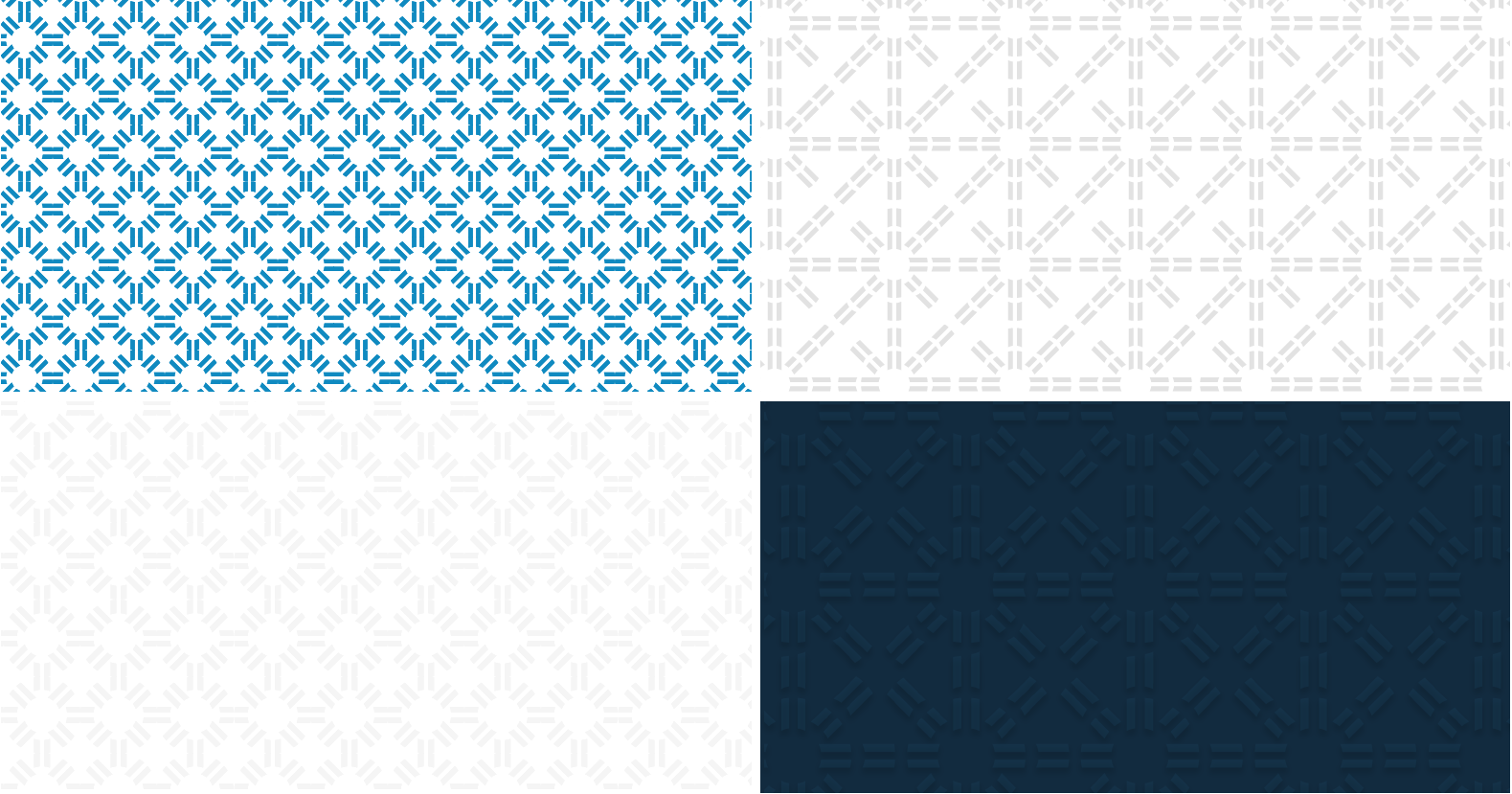
Patterns derived from our core symbol
The new brand identity incorporates a modular pattern system derived directly from the logo symbol. These patterns can be used in both subtle and expressive applications — from digital backgrounds to printed materials — helping create a unified visual environment across Czech-BioImaging.
Where will the new identity appear?
The updated identity will gradually be introduced across:
Our goal is to make the transition smooth and ensure that all materials reflect the same high standard of clarity, professionalism, and visual cohesion.
A brand that represents our community
This new visual identity underscores our long-standing commitment to open access, expert support, and high-quality imaging services. It also reflects the collaborative nature of our infrastructure — shaped by the work of our facilities, the contributions of our staff, and the creativity and needs of our users.
We look forward to presenting Czech-BioImaging with an identity that supports our mission and strengthens the visibility of the imaging community in the Czech Republic and beyond.
Next Page »« Previous Page
