30 November – 4 December 2026
Institute of Molecular Genetics, Vídeňská 1083, 142 20, Prague
The course is designed for beginners and intermediate users of transmission electron microscopes. Upon completion of the course, participants will understand the principles of transmission electron microscope design and operation, and will receive extensive hands-on experience with transmission electron microscopes of varying complexity. Participants are encouraged to bring their own samples on TEM grids for imaging. While the focus will be on routine TEM operation, a demonstration of the cryoTEM workflow and a brief overview of sample preparation methods are also included in the program.
The course will be taught in English.



8-9 September 2026
Krč campus, Vídeňská 1083, Prague 4, Czech Republic (hybrid)
This two-day introductory course, co-organized by three Czech-BioImaging Core Facilities – the Microscopy Service Centre IEM CAS, Core Facility Magnetic Resonance ISI CAS, and IPHYS Bioimaging Facility – focuses on how preclinical imaging facilities can actively support and enhance animal-based research, from study planning to data acquisition. Aimed at researchers and technical staff new to preclinical models or imaging workflows, the course introduces essential principles of proper and ethical animal handling in the specific context of facility-based imaging experiments.
Participants will learn how imaging facilities contribute to animal welfare, regulatory compliance, and experimental quality by providing expertise in animal preparation, handling during imaging procedures, and protocol optimization. The course emphasizes the value of early collaboration with facility staff to reduce experimental variability, improve reproducibility, and ensure efficient use of animal models.
The course will introduce several methods used in multimodal preclinical imaging, including in vivo two-photon microscopy, in vivo multiphoton-based label-free techniques (SHG, THG, CARS), and magnetic resonance imaging (MRI) of small rodents, illustrated through real-world examples and case studies.
The course is designed for researchers (PhD candidates, Postdocs, Principal Investigators, etc.) with a serious interest in integrating in vivo imaging into their projects.
two-photon fluorescent microscopy (functional calcium/voltage imaging, structural imaging), second harmonic generation (SHG), third harmonic generation (THG), coherent anti-Stokes Raman spectroscopy (CARS), high field magnetic resonance perfusion imaging (MRI), fluorescence lifetime imaging microscopy (FLIM)
Course fee: 40 EUR
Registration deadline: 14. 8. 2026
Payment deadline: 15. 8. 2026


1-4 December 2026
To Be Announced
This comprehensive course is designed specifically for biologists to master cutting-edge image analysis software. It covers both the theory of image processing and its practical applications, focusing on precise and efficient image quantification, segmentation, and visualization techniques. Participants will gain hands-on experience with industry-leading commercial and open-source tools and elevate their data analysis skills to the next level.
The course also introduces the FAIR principles for scientific data and demonstrates effective strategies for data management using the OMERO database.
The course is organised by the Cellular Imaging Core Facility in collaboration with the Faculty of Informatics, Masaryk University.

8-11 September 2026
CEITEC MU, University Campus, Building C02, CELLIM CF, Brno
The Advanced Light Microscopy Methods is an advanced course for students, postdocs, technicians, and other laboratory staff in biology and biomedical science who require specialised microscopy methods for their experiments. The course consists of theoretical lectures (25%) and practical hands-on sessions on selected microscopes (50%) and image processing software (25%).
Overall, this course provides an overview of core principles behind super-resolution, FLIM-FRET and mesoscopic imaging methods, together with corresponding essential image processing steps. Participants will learn theoretical principles behind large sample imaging, super-resolution methods like Airyscan, Optical Reassignment, Structured Illumination and Single Molecule Localisation, as well as Fluorescence Lifetime and Förster Resonance Energy Transfer Microscopy.
A dedicated practical session will demonstrate the learned principles directly on real microscope acquisitions. Participants will gain skills needed to operate selected microscopes and experience how to setup advanced experimental workflow, and perform the most common troubleshooting.
The workshop concludes with an overview of related workflows in image processing and analysis utilising related software (ZEN, LAS X). Finally, the participants will receive comprehensive guidelines for advanced analysis and image visualisation in Imaris.
Practical training will be conducted on the following systems:
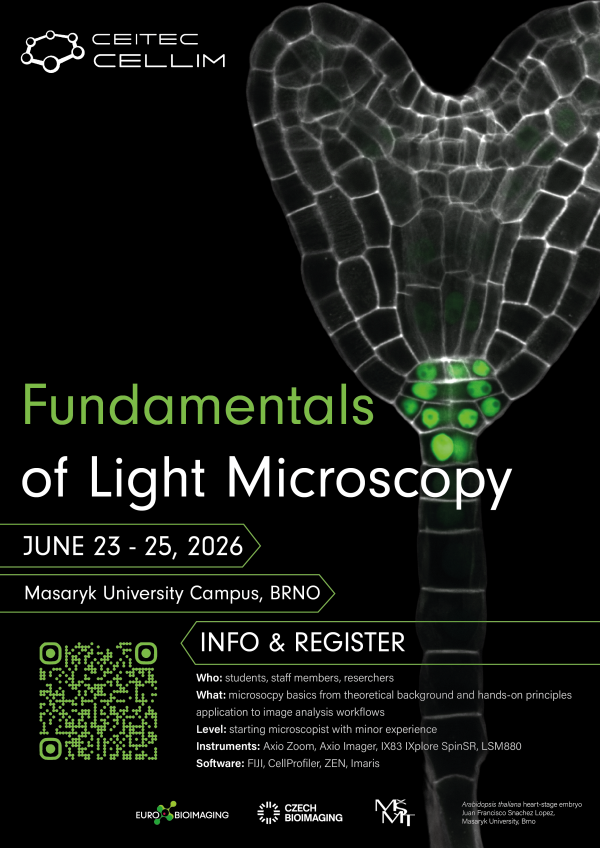
23-25 June 2026
CEITEC MU, University Campus, Building C02, CELLIM CF, Brno
The Fundamentals of Light Microscopy is a basic course for students, postdocs, technicians, and other laboratory staff in biology and biomedical science who require widefield and/or confocal microscopy for their experiments. The course consists of theoretical lectures (50%) and practical hands-on session on selected microscopes and software (50%).
Overall, this course provides an introduction to the fundamentals of light microscopy, with a focus on understanding how images are formed and how acquisition choices affect resolution, contrast, and quantitative image quality. Participants will learn core principles including diffraction and the point spread function, numerical aperture and refractive index effects, sampling and digitization, detector characteristics and noise, and the conceptual differences between major imaging modalities such as widefield, confocal, super-resolution and light sheet microscopy.
A dedicated practical session will demonstrate the learned principles directly on real microscope acquisitions. Participants will gain skills needed to operate selected microscopes and experience how changing parameters influences contrast, optical sectioning, signal-to-noise ratio, and artifacts.
Finally, the workshop concludes with an introduction to basic workflows in image processing and analysis, including foundational steps for viewing, correcting, and preparing microscopy datasets for downstream quantification using open-source and commercial tools (FIJI/ImageJ, CellProfiler, ZEN, Imaris).
8 – 13 October 2026
First Medical Faculty, Charles University, CAPI, Salmovská 3, 120 00 Prague
Language: English
The event “Imaging Horizons”, organized by the Center for Advanced Preclinical Imaging (CAPI) at the First Faculty of Medicine, Charles University, aims to raise awareness of modern imaging methods and their importance for preclinical research. The goal is to present the wide range of technologies available at CAPI, including MRI, PET, CT, ultrasound, photoacoustic imaging, in vivo fluorescence, and magnetic particle imaging, and to demonstrate their practical applications in medicine and biomedicine.
The program combines a 2.5-hour block of expert lectures featuring real data from CAPI projects, followed by a networking session to foster collaboration between participants and experts. The lecture block will be streamed online to enable interactive engagement with a broader audience and increase the event’s reach. Another part of the program consists of guided tours of CAPI laboratories, organized in four sessions (two on the day of the lectures and two on a separate day close to the event date). Participants will be divided into small groups and will learn about measurement principles, sample preparation, instrument demonstrations, and typical data outputs.
The event targets advanced master’s students, PhD candidates, and early-career researchers from medicine, biomedicine, biology, physics, chemistry, and related disciplines. A unique feature of CAPI is its multimodal approach, in-house technology development, and integration into the European Euro-BioImaging infrastructure, which facilitates connections with the international scientific community and supports new collaborations.
Link coming soon

2 – 4 December 2026
Institute of Experimental Medicine, Vídeňská 1083, 142 20, Prague
This course is aimed at participants familiar with the basics of image processing, who want to automate their analysis using scripting. We will work with ImageJ macro language and use it to create a workflow for segmentation, batch processing and analysis of fluorescence microscopy images. In the second part of the course, we will introduce Cellpose, a machine learning algorithm for cell segmentation, demonstrate its use through command line and ImageJ plugin, and incorporate its output into the ImageJ workflow.
No previous programming knowledge is expected, and the course will therefore also briefly introduce basic programming concepts, such as variables, for loops and conditionals (if-else statements). Most of the sessions will be hands-on, and the last day will be dedicated to individual consultations of user projects and questions.
The course is primarily aimed at participants with some experience with ImageJ and basic understanding of bioimage analysis. We expect no or minimal knowledge of programming.
Participants are required to bring their own laptop for hands-on exercises. If you do not have access to a laptop, please inform us in advance, and we will provide you with one. Prior to the event, you will receive a list of the required software programs to install for the course.
Please make sure that all required software is installed and works prior to the workshop, especially if your laptop runs macOS or Linux (we work mostly with Windows and are therefore not as familiar with the other operating systems, therefore troubleshooting during the course is difficult).
28 – 29 May 2026
Institute of Experimental Medicine, Vídeňská 1083, 142 20, Prague
This course introduces participants to the fundamentals of volumetric and time-lapse microscopy data processing, with a focus on visualization, segmentation, and quantitative analysis. The first day offers a hands-on introduction to Imaris, covering its core modules (Spots, Surfaces, and Filaments). The second day builds on this and includes a broader discussion on big data challenges, alternative 3D processing tools in ImageJ/Fiji, and options for data storage, sharing, and reuse (FAIR data).
The course is organized in hybrid form and it is possible to join either in-person or online.
Imaris is the main software used, and the course is designed to provide a comprehensive overview of its capabilities. As such, it is primarily aimed at participants with no or limited experience. However, advanced features and exercises will also be included to benefit those already familiar with the software.
24 – 25 November 2026
Building Da I. IPHYS BIF, Krc Campus, Videnska 1083, Prague
Course fee is 40 Euros
The two-day course consists of lectures and hands-on sessions which will demonstrate basic mechanical testing methods such as atomic force microscopy, Brillouin microscopy, indentation test or uniaxial and biaxial tensile test. The tests will be introduced in practical sessions together with their biological and methodological relevance. To relate the mechanical tests to the most significant biomechanical structures such as collagen fibers, elastic fibers or fat, the participants will be taught how to link these mechanical properties and microscopy images. Due to the minimum sample alternations, the images are acquired using label-free techniques, for instance, pSHG, THG or CARS or confocal reflectance microscopy.
1 – 4 December 2026
Institute of Scientific Instruments of the Czech Academy of Sciences, Královopolská 147, Brno, Czech Republic
The three/four-day (TBD) course organized by the ISI EM team at the Institute of Scientific Instruments of the Czech Academy of Sciences will offer students and young researchers in the fields of life sciences and soft materials both theoretical and practical training in electron microscopy and Raman spectroscopy. Participants will gain hands-on experience with SEM and FIB-SEM microscopes, biological sample preparation, EDX analysis, and cryo-SEM workflow. The program will combine introductory lectures with intensive hands-on sessions in small groups and will be complemented by a social evening and discussion sessions at the end of each day.
Link coming soon
20 – 24 April 2026
First Medical Faculty, Charles University, CAPI, Salmovská 3, 120 00 Prague
Language: Czech
Course for acquiring qualification and professional competence (designing experiments and experimental projects) in the field of experimental animal use according of Act No. 246/1992 On the Protection of Animals Against Cruelty.
Link coming soon
9 – 13 March 2026
First Medical Faculty, Charles University, CAPI, Salmovská 3, 120 00 Prague
Language: English
Course for acquiring qualification and professional competence (designing experiments and experimental projects) in the field of experimental animal use according of Act No. 246/1992 On the Protection of Animals Against Cruelty.
Link coming soon