
Understanding the three-dimensional organization of proteins is essential for explaining how they function in living cells. A new study by Štěpánka Bílá, Dominik Pinkas, Vitaly Polovinkin and colleagues, published in Nature Communications presents IncelluloED, an innovative workflow that enables researchers to determine high-resolution protein structures directly from a single crystal inside a single cell. The study is the outcome of close collaboration between a user and the staff of the Electron Microscopy Core Facility at the Institute of Molecular Genetics of the Czech Academy of Sciences (IMG CAS), a core facility of the Czech-BioImaging research infrastructure. Dominik Pinkas, one of the study’s authors, is a staff member of the facility, whose expertise and equipment were instrumental in developing and implementing the cryogenic electron microscopy workflow.
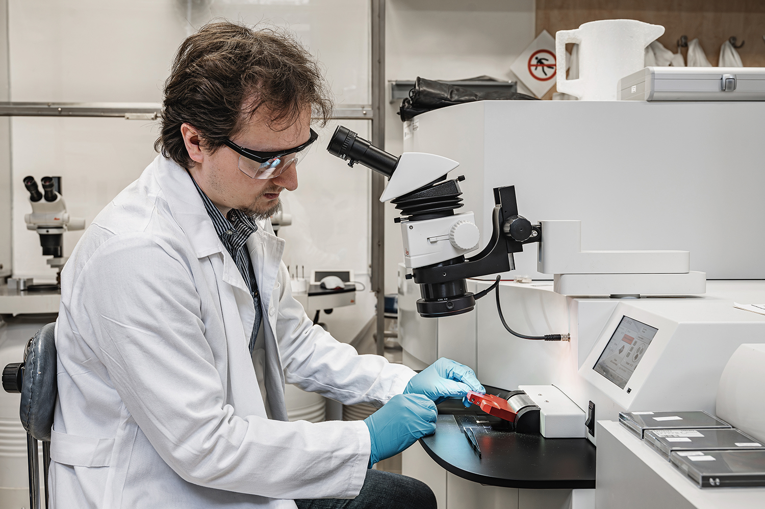
Dominik Pinkas, Electron Microscopy Core Facility, Institute of Molecular Genetics of the Czech Academy of Sciences. Photo: Czech-BioImaging archive.
IncelluloED determines high-resolution protein structures from a single crystal inside a single cell
Protein crystallization inside living cells offers an alternative route to structural biology that avoids protein purification and can preserve proteins in a quasi-native cellular environment. However, intracellular crystallization is often inefficient, and only a small fraction of cells may contain suitable crystals. Conventional serial X-ray crystallography therefore requires tens of thousands of crystal-containing cells, limiting studies of proteins that crystallize only rarely.
The newly developed IncelluloED workflow combines intracellular protein crystallization with in situ three-dimensional electron diffraction. The method enables high-resolution structure determination from a single protein crystal located inside a single cell. It builds on standard cryogenic electron-microscopy technologies, including cryo-light microscopy, correlative imaging, cryo-focused ion-beam milling and electron diffraction in a cryo-transmission electron microscope.
As a proof of concept, the researchers studied HEX-1 protein from the filamentous fungus Magnaporthe grisea. The protein was expressed in insect cells, where it formed intracellular microcrystals approximately 4-15 µm in size. Crystal-containing cells were deposited on transmission electron microscopy grids and vitrified by plunge freezing.
The frozen grids were first examined by cryo-light microscopy. Co-expression of a fluorescent marker made the crystals visible as sharply defined regions lacking fluorescence. Combined reflected light and fluorescence imaging provided the three-dimensional coordinates of selected crystals relative to the sample surface.
These coordinates were transferred to a cryo-FIB/SEM microscope by correlating the light-microscopy and electron-microscopy images. The targeted cells were then milled with a focused ion beam to produce electron-transparent lamellae approximately 250–300 nm thick. The lamellae were transferred to a cryo-TEM, where continuous-rotation electron-diffraction data were collected directly from the intracellular crystal. The complete workflow proceeds from crystal detection and plunge freezing through correlative localization, targeted lamella preparation and final diffraction-data acquisition.
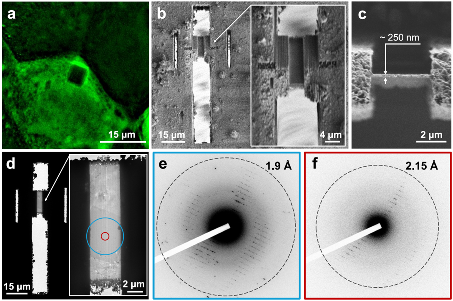
Example of a prepared lamella containing an in cellulo MgHEX-1 crystal and its electron diffraction (ED) (a) shows HEX-1 crystal in negative fluorescence imaging, (b) and (c) show lamella preparation through the same crystal. The crystal is clearly visible in the TEM in (d) while (e) and (f) show diffraction patterns obtained from blue and yellow areas of the crystal, respectively. (Nature Communications 17, 2109 (2026))
High-resolution structure from a single intracellular crystal
Using a crystal volume of approximately 1.6 µm3, IncelluloED produced the previously unknown MgHEX-1 structure at 1.9 Å resolution. A second dataset collected from only about 0.8 µm3 yielded a structure at 2.2 Å resolution. Both structures contained detailed information sufficient for modelling most of the protein backbone and side chains.
1 crystal
intracellular
⟶
1 cell
single-cell sample
⟶
1.9 Å
resolution
For comparison, serial synchrotron X-ray crystallography achieved a resolution of 1.8 Å but required data from 62,496 intracellular crystals, representing a combined crystal volume of approximately 11 million µm3. The electron-diffraction and X-ray structures showed no major differences in the overall protein conformation.
IncelluloED therefore reduces the required sample volume by approximately seven orders of magnitude while retaining structural information comparable to established serial X-ray methods. Because the workflow uses instrumentation already available in many cryo-EM facilities, it could make high-resolution analysis of rare intracellular crystals accessible without requiring synchrotron or X-ray free-electron-laser experiments.
Towards a “single-cell structural laboratory”
The method remains to be validated on a broader range of proteins and crystal symmetries. Nevertheless, it introduces a practical route for studying proteins that crystallize inefficiently, detecting structural heterogeneity that would otherwise be averaged across thousands of crystals, and investigating intracellular protein structures within their cellular environment. It represents an important step towards a “single-cell structural laboratory.”
Article: Bílá, Š., Pinkas, D., Khakurel, K. et al. Single-cell structural biology with intracellular electron crystallography. Nat Commun 17, 2109 (2026). https://doi.org/10.1038/s41467-026-69205-6
Artistic visualisations of the IncelluloED method:
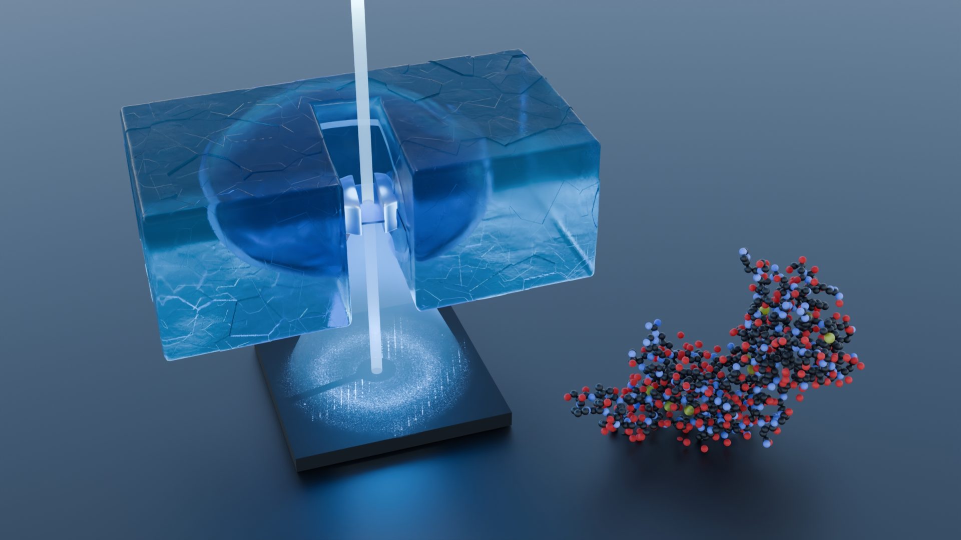
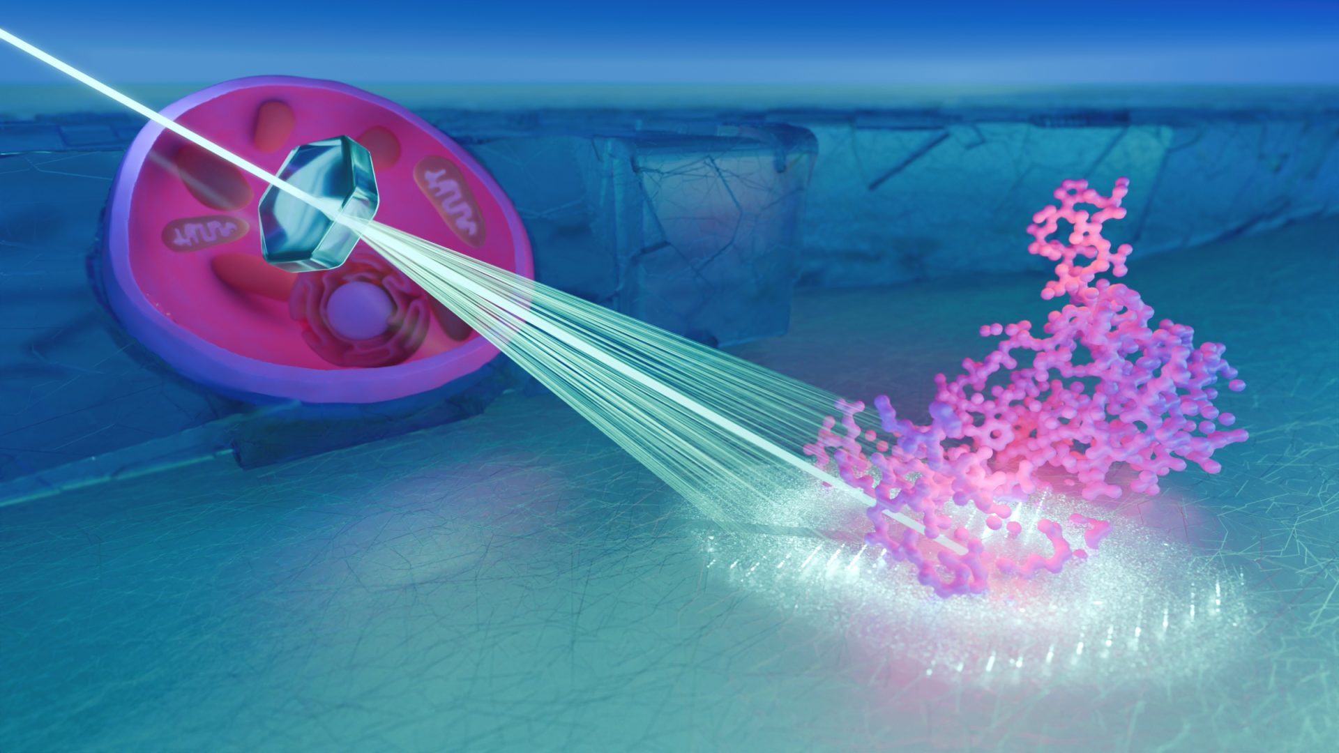
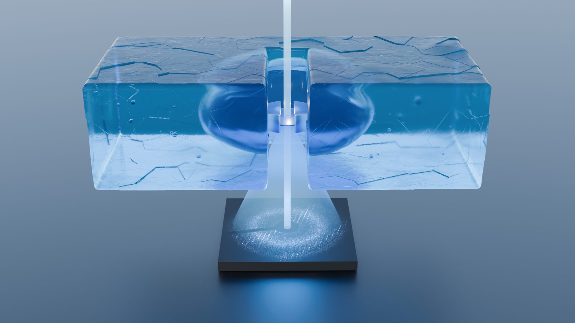
Artistic illustration of IncelluloED method by Lucas J. Martin, Max Planck Institute of Biophysics, Frankfurt am Main, Germany