
International exchange is one of the most effective ways to advance scientific expertise. Earlier this month, Czech-BioImaging welcomed Vaishali Kailaje, Senior Scientific Assistant, Facility In-charge, Digital Imaging Facility at the Advanced Centre for Treatment, Research and Education in Cancer (ACTREC), Kharghar, Navi Mumbai, India, for a two-week visit through the Global BioImaging International Job Shadowing Program.
From 1 to 12 June 2026, Vaishali visited several Czech-BioImaging facilities in Prague, gaining hands-on experience with state-of-the-art imaging technologies while working alongside imaging specialists from across our network. Her programme combined one-on-one mentoring, practical training on cutting-edge imaging platforms, and participation in the Super-Resolution in Light Microscopy Course, offering a comprehensive overview of the expertise and technologies available within Czech-BioImaging.
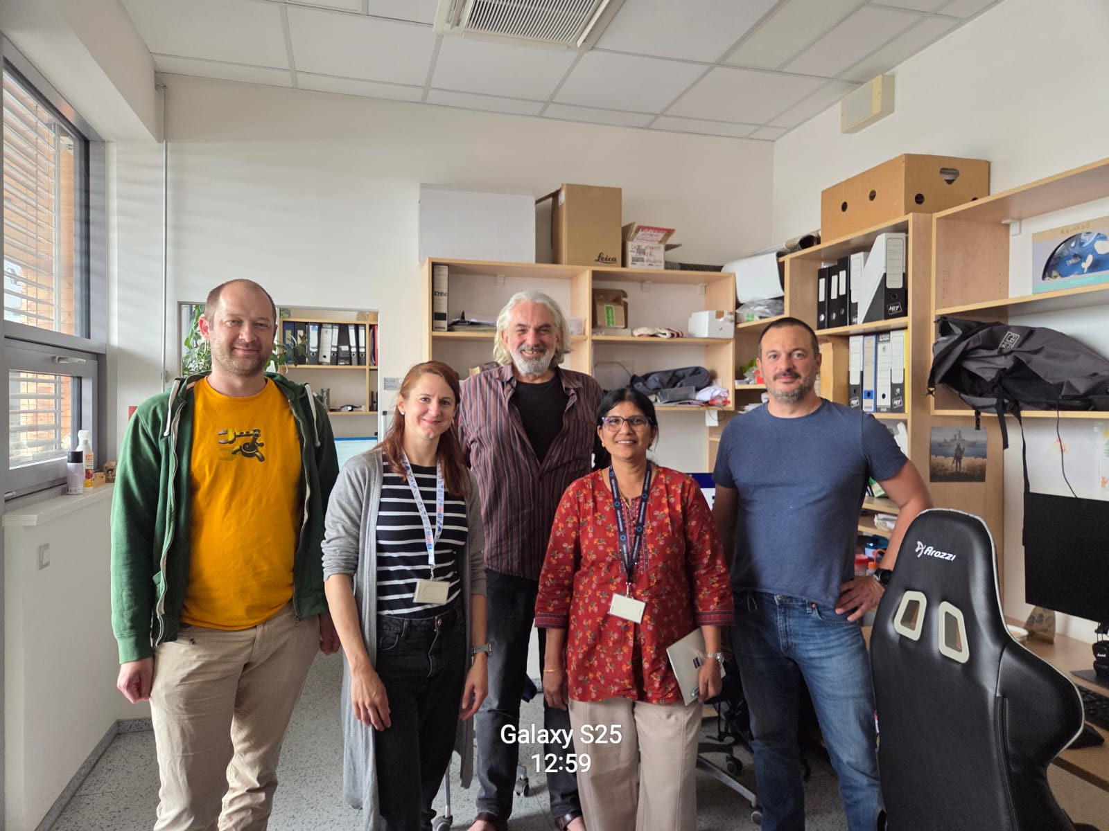
Vaishali Kailaje (second from right) with the IMG Light Microscopy Core Facility team: Jan Valečka, Michaela Blažíková, Ondrej Horváth and Ivan Novotný.
Expanding expertise in advanced imaging
Reflecting on her objectives, Vaishali explains: “The primary goal of my visit was to gain hands-on experience with fluorescence techniques such as FRET, FRAP, FLIM, and FCS, as well as super-resolution methods including STED, SIM, STORM, and PALM. I am delighted to say that this objective was fully achieved thanks to the excellent guidance and support provided by the experts and mentors I had the privilege of meeting.”
During her stay, Vaishali trained across four Czech-BioImaging facilities in Prague. At the Imaging Methods Core Facility of BIOCEV (CUNI IMCF BIOCEV), she explored label-free imaging, FLIM, FCS, lattice light-sheet microscopy and STED. At the Light Microscopy Core Facility (IMG LM), she focused on live-cell imaging, high-content screening, FLIM-FRET, TIRF and spinning disk microscopy while participating in the Super-Resolution in Light Microscopy Course. At the Institute of Physiology BioImaging Facility (IPHYS BIF), she gained experience with Brillouin microscopy alongside label-free imaging and FLIM. Her programme concluded at the Institute of Experimental Medicine Microscopy Service Centre (IEM MSC), where she explored live-cell super-resolution imaging using Airyscan, intravital imaging of mice brain and image analysis using Imaris software.
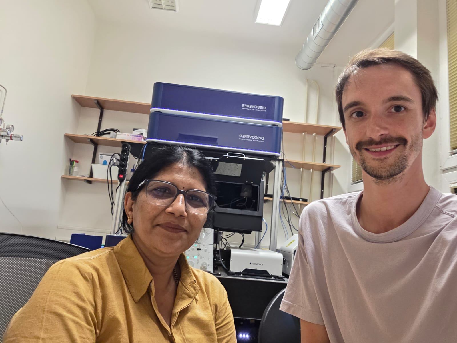
At the IPHYS BIF with Šimon Vrana in front of the Brillouin microscope.
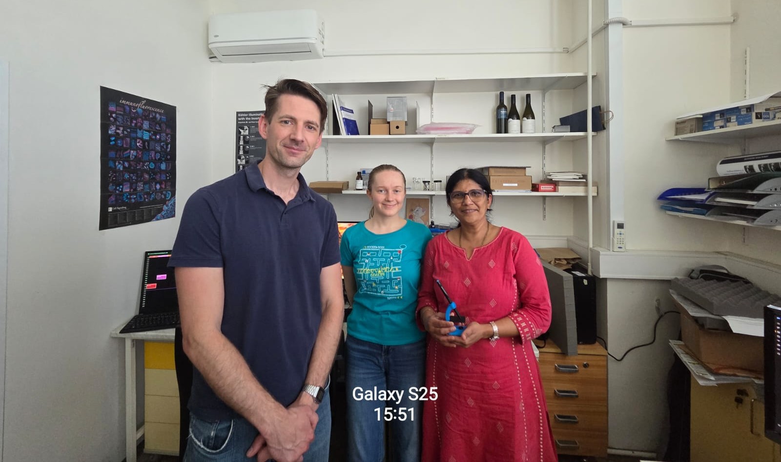
At the IEM MSC with Štěpán Kortus and Iva Švecová.
Hands-on experience with cutting-edge technologies
Throughout the programme, Vaishali received practical training on the Leica STED system, Zeiss Elyra for SIM and TIRF-SMLM, Olympus SoRa and ScanR systems, the Discoverer™ Brillouin Microscope, the NanoLive label-free imaging system, and the Akoya multiplex imaging platform. She also observed correlative cryo-electron microscopy workflows and intravital mouse brain imaging for the first time – these experiences she describes as among the highlights of her visit.
Working closely with imaging specialists across the Czech-BioImaging network, Vaishali particularly appreciated the openness with which the teams shared their expertise, workflows, and protocols. A highlight of the programme was the Super-Resolution in Light Microscopy Course, whose combination of lectures and extensive practical sessions significantly deepened her understanding of super-resolution imaging and the optimization of acquisition parameters. She expressed special thanks to Ivan Novotný for enabling her participation in the course and for creating an excellent learning environment.
Building connections for the future
Beyond developing technical skills, the visit created valuable opportunities to build professional relationships across the Czech-BioImaging community. These new connections will continue beyond the job shadowing, providing a network of colleagues she can turn to for advice and future collaboration. The knowledge and experience gained during the programme will support the further development of imaging applications and instrumentation at ACTREC, strengthening the facility’s ability to support researchers interested in advanced microscopy techniques.
Looking back on her two weeks with Czech-BioImaging, Vaishali says the experience extended far beyond learning new microscopy techniques. “My visit was much more than a training course—it was a journey that fostered knowledge, friendships, and future collaborations.”
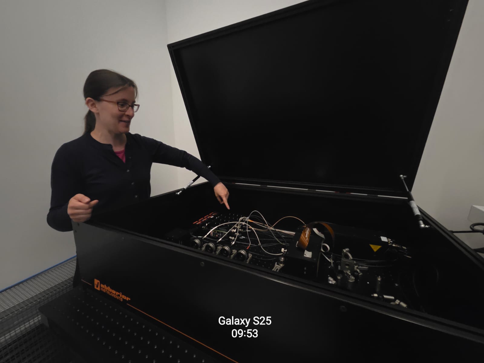
Marie Olšinová showing the Abberior Instruments STED microscope at the CUNI IMCF BIOCEV.
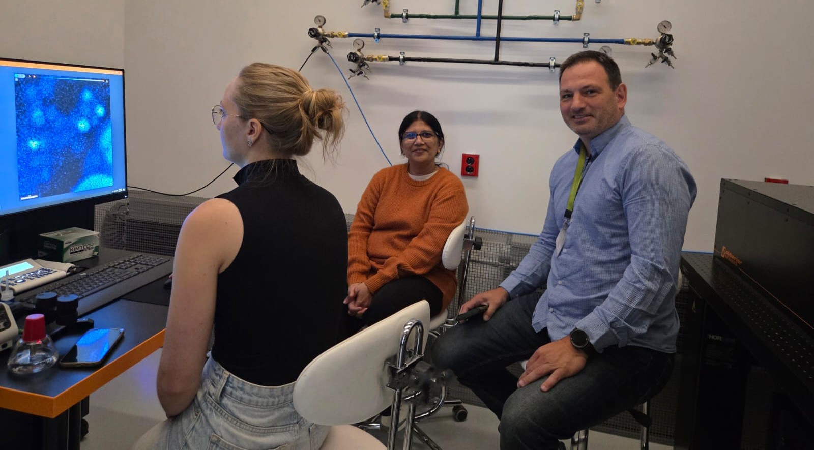
Kateřina Paldusová from the J. Heyrovský Institute of Physical Chemistry, a Czech-BioImaging user, reviewing STED microscopy images with Aleš Benda.
Strengthening the global bioimaging community
Vaishali’s visit was made possible through the Global BioImaging International Job Shadowing Program, which supports international exchange between imaging facilities worldwide. She expressed her sincere gratitude to the Global BioImaging community for the travel grant that made the visit possible, to India BioImaging for introducing her to the Global BioImaging platform, and to the Czech-BioImaging network for providing such an outstanding learning opportunity. She noted that the experience had been immensely enriching, both professionally and personally, and that the knowledge, friendships, and professional connections established during her stay would remain invaluable throughout her career.
International exchanges like this demonstrate the value of sharing expertise across imaging communities. We were delighted to welcome Vaishali to the Czech-BioImaging network and look forward to seeing the new collaborations and scientific advances that grow from this experience.