
Published in Nature Communications, the study by Gui et al. uncovers an unexpected role of the protein MCL1 in driving tumour metabolism, challenging a long-standing view of its function in cancer. The work was supported by advanced in vivo PET imaging performed at the Center for Advanced Preclinical Imaging, Charles University, a Czech-BioImaging and Euro-BioImaging facility. We spoke with the paper’s corresponding author, Dr. Mohamed Elgendy (Medical Clinic I, University Hospital Carl Gustav Carus, Technische Universität Dresden, Dresden, Germany), about the significance of the findings, their potential clinical impact, and the role of imaging in the study.

For years, cancer researchers have viewed the protein MCL1 primarily as a protective shield for tumors—a classical “anti-apoptotic” brake that prevents cancer cells from self-destructing. However, your new study shattered this view. Could you explain how MCL1 acts as more than just a shield, and why your findings represent such a major paradigm shift?
MCL1 is one of the most highly expressed proteins in several types of tumors and is an anti-apoptotic member of the Bcl-2 family, a family of proteins that are typically viewed as effectors crucially deciding about cell death or survival downstream of DNA damage, ER stress, metabolic crisis, etc.
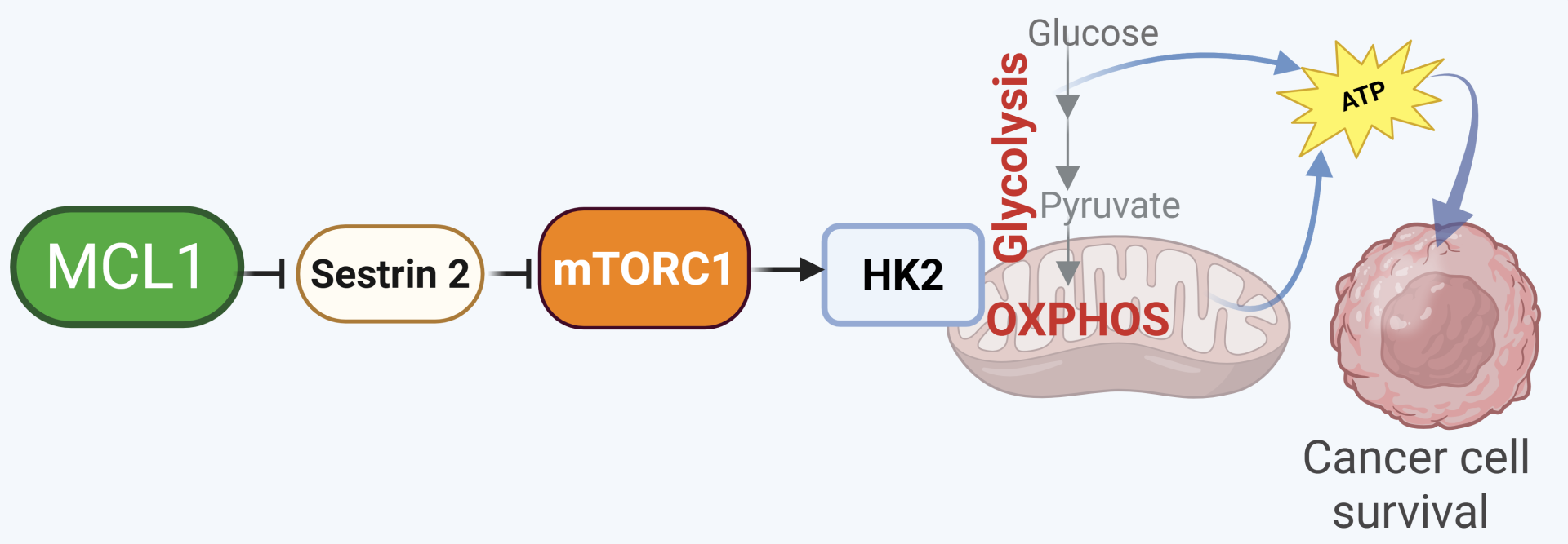
The study thus represents a major paradigm shift: It positions – for the first time to the best of our knowledge – MCL1 as an upstream regulator that signals to a key signaling cascade (mTOR signaling), thereby challenging the classical view of MCL1 as merely a downstream effector. Moreover, the tumor-promoting role of MCL1 has so far been largely attributed to its anti-apoptotic function. Here, we show that the novel functions of MCL1 in controlling mTORC1 signaling and subsequently in regulating bioenergetics contribute to tumor promotion by MCL1.
The ground-breaking nature of this discovery is illustrated by the fact that it was selected among the best 50 Cancer articles in Editor’s highlight and received high visibility (Top 1% of the 349,562 tracked articles of a similar age in all journals and top 5% in Nature Comm. https://www.nature.com/articles/s41467-025-66831-4/metrics).

This completely changes our understanding of tumor metabolism. But how do these laboratory findings translate to real-world patient care, specifically for patients fighting melanoma or AML?
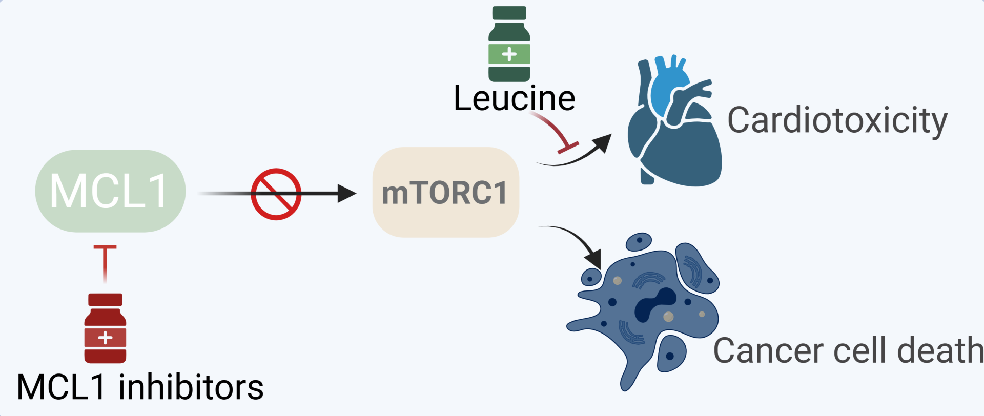
From a clinical preservative, the study has an immediate clinical relevance. Targeting MCL1 is emerging as a promising therapeutic strategy with several inhibitors under clinical development. Our findings show that MCL1 inhibitors act to inhibit mTOR signaling. mTOR inhibitors (mTORi) are used clinically in cancer therapy and therefore the unexpected and unintended property of MCL1 inhibitors on mTOR inhibition we show here is very relevant clinically.
Moreover, several clinical trials testing MCL1 inhibitors have been halted or discontinued due to reported cardiotoxicity. Importantly, in the light of our novel insights, we identify a crucial mechanism of the cardiotoxicity of MCL1 inhibitors and finally devise a dietary approach to dramatically ameliorate such cardiotoxicity. This is likely to be vital for further advancing the use of those inhibitors in clinical testing. Additionally, our data establish MCL1-mTORC1-HK2 as an important prognostic axis in melanoma and AML.
Your study relied on advanced imaging approaches to understand how MCL1 influences tumor metabolism in vivo. Could you explain how imaging technologies helped validate your findings?
Our in-vitro data indicated that depletion or inhibition of MCL1 leads to a decline in glucose metabolism and glycolysis. As metabolism of cancer cells in vitro may differ from that of tumors in vivo, we aimed to assess the role of MCL1 in regulating tumor metabolism in vivo
We validated our findings in vivo through collaboration with the group of Prof. Ludek Sefc and the team of the Center for Advanced Preclinical Imaging (CAPI) at the Charles University in Prague, which is supported by Euro-BioImaging and Czech-BioImaging funding. We exploited the advanced imaging technique of in vivo 18-fluorodeoxyglucose positron emission computed tomography (18FDG PET), an imaging technique that assesses the uptake of radio-labeled 18FDG and is routinely used to monitor glucose metabolism in tumors 18FDG-PET scan. This technique allows for the direct quantification of glucose uptake, an indicative readout of glucose metabolism and glycolysis.

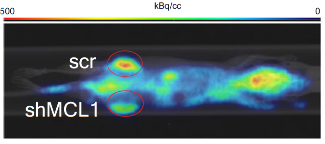
In-vivo 18FDG-PET scan was performed on tumor subcutaneous xenograft derived from melanoma cells that were either depleted or not of MCL1. This analysis further confirmed the findings obtained from the CHL-1 model. We have further normalized the data according to injected FDG, quantified FDG uptake in internal organs (heart, brain and kidney), and quantified PET imaging calculating SUVmax alongside gamma counting.
Nod skid gamma (NSG) mice received single subcutaneous flank injections with melanoma transduced with either scrambled shRNA or doxycycline -inducible shRNA against MCL1 on both flanks. Once tumors were established, mice were given drinking water containing 1 mg ml-1 doxycycline and 1% sucrose for induction of MCL1 shRNA expression in vivo for five additional days. Mice were fasted overnight (14-18h), weighed and heated prior to intravenous injection of 18FDG through the tail vein (activity approx. 6 MBq per mouse). Animals were placed into heated induction chamber under anesthesia (1.5% isoflurane) for 40min uptake. The 10min CT-PET imaging (Albira, Bruker, Germany) under anesthesia (1.5% isoflurane) was performed running PET measurement (10min, PET single, offset 35mm) & CT (2x 8min, 125mm LOW 400kV/400µA, offset 35mm). Tumors were isolated, weighed, and their size determined using digital caliper. The 18FDG activity in blood samples and extracted tumors was measured using a 2480 Wizard2® Automatic Gamma Counter (PerkinElmer, USA), and corrected for the half-life decay. CT-PET image analysis and co-registration were carried out using PMOD analysis software (PMOD Technologies LLC; Switzerland). Consistent with the in-vitro results, MCL1 depletion in tumors led to decline in 18FDG signal as compared to control tumors, suggestive of inhibition of glucose metabolism in MCL1-depleted tumors. Of note, at the time of tumor isolation there was no significant difference in tumor size between both groups of tumors. Establishing the control and MCL1-deplted tumors on both flanks of mice controlled for the inter-mouse and inter-organ variabilities of 18FDG uptake as it allowed comparison of two tumors established in the same mouse.
Original Nature Communications article: Gui, W., Paral, P., Dhamija, B. et al. MCL1 modulates mTORC1 signaling to promote bioenergetics and tumorigenesis. Nat Commun 16, 10841 (2025). DOI: 10.1038/s41467-025-66831-4
Authors:
Markéta Schmidt Černohorská, Euro-BioImaging Scientific Ambassador
Mohamed Elgendy, Medical Clinic I, University Hospital Carl Gustav Carus, Technische Universität Dresden, Dresden, Germany
Daniela Klimešová, Czech-BioImaging Project Manager